Atopic eczema and infected eczema
The commonest childhood skin disease - in the ED the job is to spot the infected flare and to fix the treatment that is not working.
Red flags
- Monomorphic punched-out erosions, or a painful rather than itchy flare - eczema herpeticum, needing aciclovir without waiting for swabs
- Fever, systemic upset, or rapidly spreading infection
- Periocular involvement in a suspected herpetic flare - same-day ophthalmology and dermatology
- Failure to thrive, or severe eczema in an infant not responding to appropriate treatment
- Erythroderma - near-total skin involvement is a medical emergency in its own right
Recognise
Ill-defined red scaly itchy patches, weeping and crusted when acute, lichenified and thickened when chronic, in a flexural distribution.
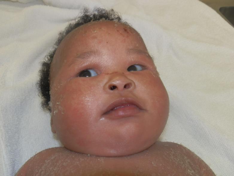
Erythema is the standard description of eczema and it is frequently absent in brown and black skin, where active inflammation appears violaceous, grey, dark brown or simply as an ashen change - which is why eczema severity is systematically under-scored in darker skin and children are under-treated as a result. Two pattern differences also matter: follicular or papular eczema, with prominent goosepimple-like papules around hair follicles, and a more extensor than flexural distribution. Sarkar and Verma's review of atopic dermatitis in skin of colour describes atypical papular and follicular morphology with more pronounced lichenification, reported particularly in patients of Asian ethnicity; the extensor distribution is widely taught but is not stated in that review. Either way these patterns are often not recognised as eczema at all. Judge severity on lichenification, excoriation, warmth and the child's sleep and scratching rather than on redness. Post-inflammatory hypopigmentation and hyperpigmentation are prominent and, in that review, persist long after the active inflammation subsides, causing significant psychosocial distress. NICE CG57 makes the same point in its own words - atopic eczema may temporarily cause the skin to become lighter or darker. They persist for months and are a major source of distress - explain that they are not scars and not steroid damage.
- Itch is essential to the diagnosis - eczema that does not itch is probably not eczema
- Personal or family history of atopy
- Lichenification and excoriation mark chronicity and severity
- Infected eczema: weeping, golden crusting, pustules, rapid worsening, or a flare that stops responding
- Eczema herpeticum: monomorphic punched-out erosions, pain rather than itch, and systemic upset - a different emergency
Distribution
Photographs
Mimics
- Eczema herpeticum — The complication that must never be missed - it is treated as an emergency, not as a flare
- Impetigo — Bacterial infection of eczema is common and the two frequently coexist
- Nappy rash, candida, and patterns that should worry you — In infants, irritant and candidal nappy dermatitis is distinguished by involvement or sparing of the skin folds
- Erythroderma — Eczema is a leading cause of erythroderma when it becomes near-total
- Acute urticaria — Acute urticaria is itchy but the weals move within hours and leave no scale
- Pompholyx — Pompholyx is a vesicular pattern of eczema on the palms and soles, and many patients have atopic eczema elsewhere
What to do in the ED
- Establish first whether this is a plain flare, a bacterially infected flare, or eczema herpeticum - the three have different urgencies
- Emollients generously and frequently, and a topical corticosteroid of potency matched to severity and site, per NICE CG57's stepped approach
- Check what they are actually using and how much. Under-use of emollient and fear of topical steroid are the usual reasons a flare reaches the ED, and correcting that is often the whole intervention
- Treat bacterial infection with antibiotics where there is crusting, weeping or pustulation with systemic features
- Start aciclovir if eczema herpeticum is suspected - do not wait for confirmation
- Assess sleep, school attendance and the family's coping; severe eczema is a substantial burden and warrants dermatology referral rather than repeat ED attendance
- Explain pigment change explicitly in patients with brown and black skin - this is a common reason for lost confidence in treatment
Disposition
Optimise treatment and discharge with GP follow-up; dermatology referral for severe or poorly controlled disease. Admit for eczema herpeticum with systemic upset, severe infection, or erythroderma.
Safety-netting
Local variation
Sources
- NICE CG57 - Atopic eczema in under 12s: diagnosis and management
- NICE CKS - Eczema - atopic
- Sarkar R, Verma D. Atopic Dermatitis in Skin of Colour: A Review. Indian J Dermatol 2026
- Mukwende M, Tamony P, Turner M. Mind the Gap: A handbook of clinical signs in Black and Brown skin. St George’s, University of London (2020)