Acute urticaria
Itchy weals that move, each one gone within a day - common, usually self-limiting, and dangerous only when it is the skin half of anaphylaxis.
Red flags
- Any airway, breathing or circulatory involvement is anaphylaxis, not urticaria - voice change, stridor, throat or tongue swelling, difficulty swallowing, wheeze or breathlessness; or pallor and clamminess, tachycardia, faintness, collapse or reduced consciousness. Hypotension is a late sign and is particularly late in children - do not wait for it. Persistent vomiting or abdominal pain after a food or sting, or a sense of impending doom, belongs in the same bracket. Give intramuscular adrenaline and manage per the Resuscitation Council UK algorithm; do not wait to see whether the rash settles
- A reaction already treated with adrenaline needs observation for a biphasic recurrence, not discharge on an antihistamine - CKS notes these are hard to predict
- Angio-oedema with the weals - deep swelling of the lips, tongue, face or throat - needs the angio-oedema entry and an airway assessment, not an antihistamine and discharge
- Individual lesions lasting more than 24 hours, painful or burning rather than itchy, non-blanching or palpable, or leaving bruising or pigment as they fade - CKS says to consider urticarial vasculitis, particularly with fever, malaise or arthralgia, and that needs investigation
- A systemically unwell patient - urticaria itself does not make people unwell, so look for the illness that is causing it
- Urticaria beginning within an hour of a food, drug or sting, in a patient who is now well - still counts as a possible allergic reaction and needs advice and follow-up rather than plain reassurance
Recognise
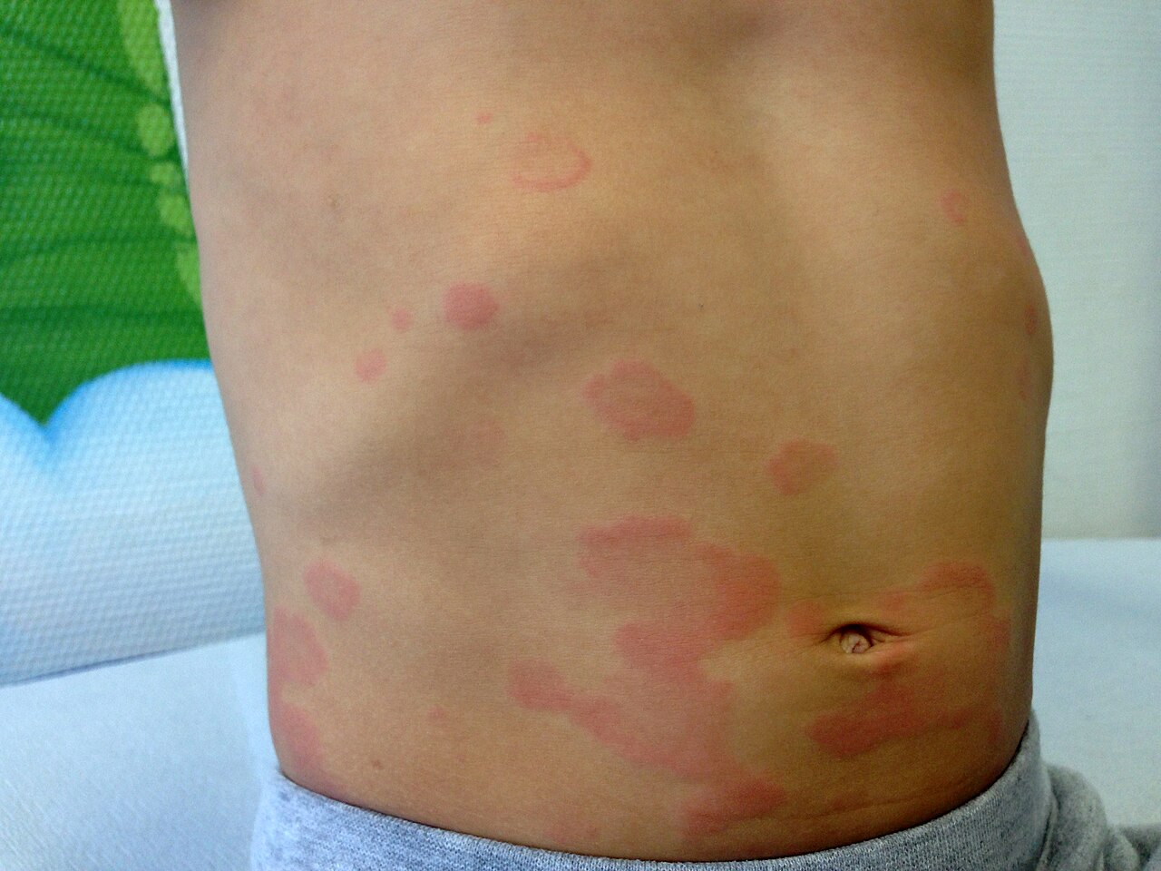
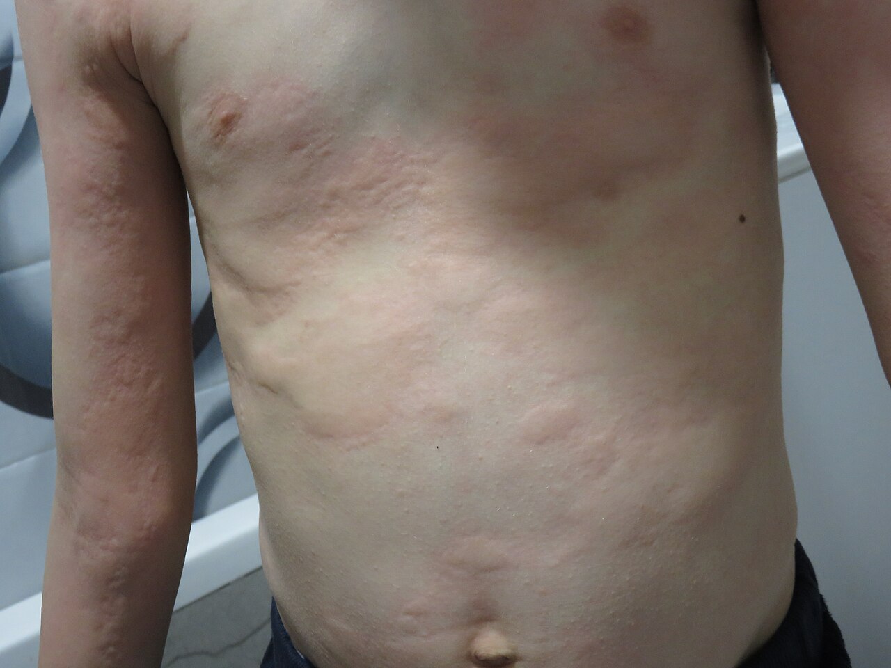
Raised pink or red weals of very variable size, often with pale centres and a surrounding flare, intensely itchy, and each individual lesion resolving within hours without a mark.
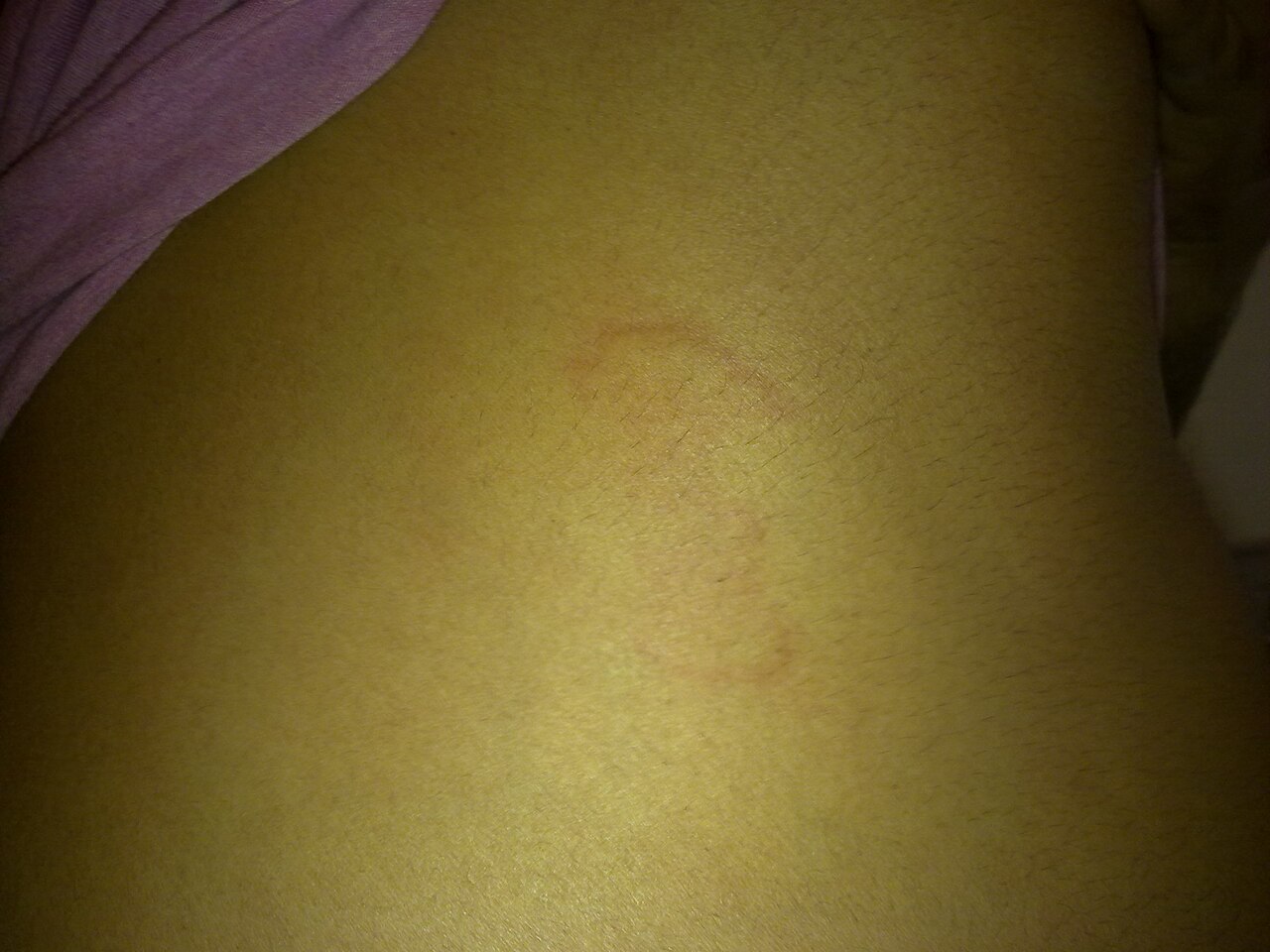
The flare - the surrounding redness that makes a weal obvious in pale skin - is frequently invisible, and the weal itself may be skin-coloured, paler than the surrounding skin, or only slightly darker, so the eruption can be missed entirely on inspection. That colour description is clinical observation rather than a cited finding; what is cited is CKS’s own wording that the central swelling may be red or white, which is the point that matters. This is a condition you diagnose with your hands and with the history. Run a hand across the skin: the weal is raised, and the change in surface contour is palpable when it is not visible. CKS's own description helps here, because it notes the central swelling may be red or white rather than assuming redness. Ask about itch, ask whether individual spots come and go within a day, and mark one with a pen to test that directly. Post-inflammatory hyperpigmentation may follow and can be mistaken for the rash persisting.
- Each individual weal lasts less than 24 hours and leaves no mark - CKS puts the fleeting nature, with skin returning to normal, at 1 to 24 hours. Mark one with a pen and re-examine: this single test does most of the diagnostic work
- Itch, or sometimes a burning sensation, is close to invariable - an eruption that does not itch is probably not urticaria
- Weals are palpably raised, of very variable size, and migratory - new ones appear as old ones fade
- Acute is under 6 weeks; chronic is 6 weeks or longer on a nearly daily basis (CKS)
- Dermographism - weals appearing in the line of a scratch - is a physical variant and is a useful bedside confirmation
- Ask about triggers: a new drug, an infection, an insect sting, a food eaten within the hour, exercise, heat or cold
Distribution
Photographs
Mimics
- Urticarial vasculitis — The distinction that matters. Ordinary weals are fleeting, itchy and leave nothing behind; vasculitic lesions outlast 24 hours, burn or hurt, and leave bruising or pigment
- Angio-oedema - allergic, ACE-inhibitor and hereditary — The deep counterpart of the same process - and the presence or absence of weals is what separates a histamine-mediated from a bradykinin-mediated mechanism
- Drug eruptions - morbilliform and fixed — A morbilliform drug rash is fixed in place for days, whereas urticarial lesions migrate within hours
- Erythema multiforme major — Target lesions are fixed, have a concentric structure and persist for days; urticarial lesions are transient and have no true target
- IgA vasculitis (Henoch-Schonlein purpura) — Early IgA vasculitis in a child can look urticarial before it becomes purpuric - palpable purpura on the buttocks and legs that does not fade is the discriminator
What to do in the ED
- Assess airway, breathing and circulation first. Urticaria is a skin diagnosis only once anaphylaxis has been excluded, and the rash is the least important part of an anaphylactic reaction
- Mark a weal with a pen and record the time. Re-examining it later is the cleanest way to demonstrate the under-24-hour behaviour, and it is the evidence the next clinician needs
- Examine by palpation as well as inspection, particularly in brown and black skin, where the weal may be felt rather than seen
- Take a focused trigger history over the preceding hours to days: drugs including over-the-counter and complementary ones, foods, stings, infections, exercise and physical stimuli
- A non-sedating antihistamine daily for up to 6 weeks, with a short course of an oral corticosteroid of up to 7 days added for severe symptoms. A one-week course of prednisolone does not need tapering. The international EAACI/GA2LEN/EuroGuiDerm guideline below is the independent reference for the stepped approach; CKS is the primary-care one
- In a child the antihistamine choice is age-dependent, and CKS says so explicitly - licensed ages differ. Cetirizine is licensed from 2 years (2.5 mg twice daily at 2 to 5 years, 5 mg twice daily at 6 to 11), and is not licensed below 2, where the BNFC gives an unlicensed dose. Fexofenadine is licensed only from 12 years. Take the dose from the BNFC against age and weight rather than prescribing as for an adult
- Where itch is disturbing sleep, a sedating antihistamine such as chlorphenamine may be added at night
- Where a specific trigger is identified, say so explicitly, document it, and give written avoidance advice - an identified trigger is the whole value of the attendance
- Where the urticaria began within an hour of a food, drug or sting, observe for a period before discharge, and make sure the patient will not be alone for the next few hours. CKS is clear that most allergic skin changes do not progress to anaphylaxis - but the ones that do, do so early
- Do not investigate acute urticaria routinely - investigations are not usually required to make the diagnosis
Disposition
Treat and discharge once anaphylaxis and angio-oedema are excluded. Acute urticaria is usually self-limiting. Refer to allergy or immunology where there is a clear and avoidable trigger with a systemic reaction, and to dermatology or immunology where it becomes chronic.
Safety-netting
Sources
- NICE CKS - Urticaria
- NICE CKS - Angio-oedema and anaphylaxis
- Lim HW et al. International Expert Consensus on Knowledge Gaps in Care for Dermatologic Disorders in Skin of Color. Int J Dermatol 2026
- NICE CG153 1.2.1.5 - erythema may be underestimated in skin types V-VI (stated in the psoriasis guideline; cited here for that general point about assessing erythema, not for this condition)
- Zuberbier T et al. The international EAACI/GA2LEN/EuroGuiDerm/APAAACI guideline for the definition, classification, diagnosis, and management of urticaria. Allergy 2022;77(3):734-766