Erythroderma
Inflammation of nearly the whole skin surface - a failure of the skin as an organ, with the physiology of a burn.
Red flags
- Hypothermia or temperature instability - the patient cannot thermoregulate and needs active warming
- Tachycardia and hypotension from transcutaneous fluid loss - this is burn physiology, not simple dehydration
- Any blistering, mucosal erosion or detachable epidermis - reconsider epidermal necrolysis
- Fever with an unwell patient - secondary bacteraemia through a failed skin barrier
Recognise
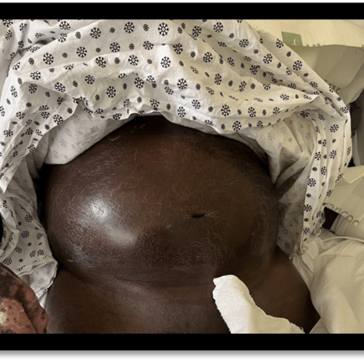
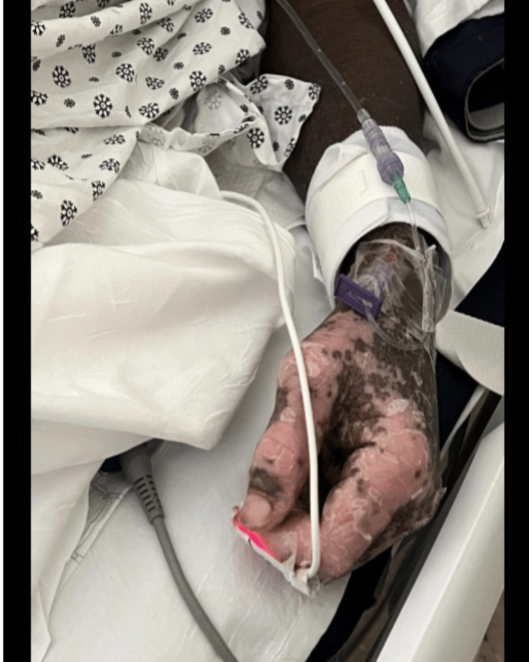
Confluent bright red skin over almost the entire surface with scaling or desquamation, often with oedema, fissuring and lichenification.
The defining redness may be entirely absent as a visual finding. Erythroderma in brown and black skin more often looks dark, violaceous, grey or simply uniformly hyperpigmented, and the diagnosis is frequently delayed for exactly this reason. Use the tone-independent signs: the skin feels hot and tight, scaling and desquamation are visible against any background, and the systemic consequences - hypothermia, tachycardia, oedema - are unaffected by pigment. Compare with an unaffected area if any remains.
- Scaling or desquamation across nearly the whole surface
- The skin feels hot and tight; the patient complains of cold and shivers
- Hypothermia or temperature instability from loss of thermoregulation
- Peripheral oedema and tachycardia from fluid and protein loss
- Lymphadenopathy is common and usually reactive
- Look for clues to the cause: nail pitting or scalp scale for psoriasis, a drug history, a long-standing eczema, or lymphoma
Distribution
Photographs
Mimics
- Stevens-Johnson syndrome / toxic epidermal necrolysis — Necrolysis also involves most of the surface but detaches epidermis and erodes mucosa; erythroderma scales without shearing
- Staphylococcal scalded skin syndrome — In a young child, generalised erythema with desquamation may be SSSS; the mucosa is spared in both, so look for the shearing
- DRESS - drug reaction with eosinophilia and systemic symptoms — DRESS can become erythrodermic; the eosinophilia and organ involvement identify it
- Toxic shock syndrome — Both give generalised erythema with later desquamation; TSS is shocked from the outset
What to do in the ED
- Treat the physiology first: warm the patient actively, measure a core temperature, and correct fluid and electrolyte losses
- Take FBC, renal and liver function, albumin, CRP, cultures if febrile, and a film if lymphoma is a consideration
- Stop any plausible culprit drug
- Emollient generously and wrap; avoid potent topical steroids until dermatology has assessed, since the cause determines treatment
- Refer to dermatology the same day and admit - this is not a discharge diagnosis at any age
- Involve burns or critical care if thermoregulation or haemodynamics are failing
Disposition
Admit with same-day dermatology. Consider burns or critical care input where thermoregulation, fluid balance or the skin barrier has failed.
Sources
- Jovic TH et al. Desquamating dermatological conditions: The burns surgeon's guide to diagnosis and management. J Plast Reconstr Aesthet Surg 2026
- Lim HW et al. International Expert Consensus on Knowledge Gaps in Care for Dermatologic Disorders in Skin of Color. Int J Dermatol 2026
- Yamamoto T. Erythroderma in the elderly. J Dermatol 2025
- Choubey S et al. Erythroderma as a presenting feature of dermatomyositis: case-based review. Rheumatol Int 2025
- NICE CG153 1.2.1.5 - erythema may be underestimated in skin types V-VI (stated in the psoriasis guideline; cited here for that general point about assessing erythema, not for this condition)