Impetigo
A superficial bacterial skin infection with a golden crust - the question worth asking is what it is growing on.
Red flags
- Systemic upset, fever or spreading erythema - cellulitis or deeper infection rather than impetigo
- Widespread bullous disease in an infant with skin tenderness - reconsider staphylococcal scalded skin syndrome
- Recurrent impetigo - look for an untreated underlying dermatosis or a colonised contact
- Failure to respond to appropriate treatment
- Peri-orbital involvement, or infection in an immunosuppressed patient
Recognise
Thin-walled vesicles or pustules that rupture to leave the characteristic golden-yellow honey-coloured crust on a red base; bullous forms show flaccid blisters leaving a collarette of scale.
The surrounding erythema is often invisible, but the golden-yellow crust itself is a colour of exudate, not of skin, and looks the same on every skin tone - so this is one of the few classic signs that transfers intact. The greater risk in brown and black skin is the opposite one: underlying eczema is frequently lichenified, follicular or hyperpigmented rather than red, so impetiginised eczema is under-recognised and treated as simple impetigo when the eczema also needs treating. Examine the surrounding skin texture. Healing leaves marked post-inflammatory pigment change, not scarring.
- Golden or honey-coloured crust - the defining sign
- Superficial, non-tender to mildly tender, and the patient is systemically well
- Spreads by scratching and autoinoculation, and between close contacts
- Bullous form: flaccid blisters that rupture easily, commoner in infants
- Ask what it is growing on - eczema, scabies, head lice or chickenpox underneath changes the treatment
Distribution
Photographs
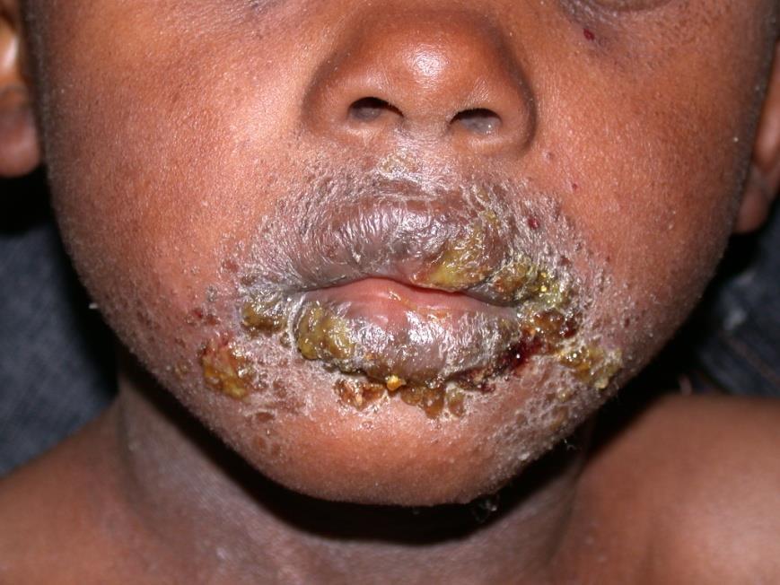
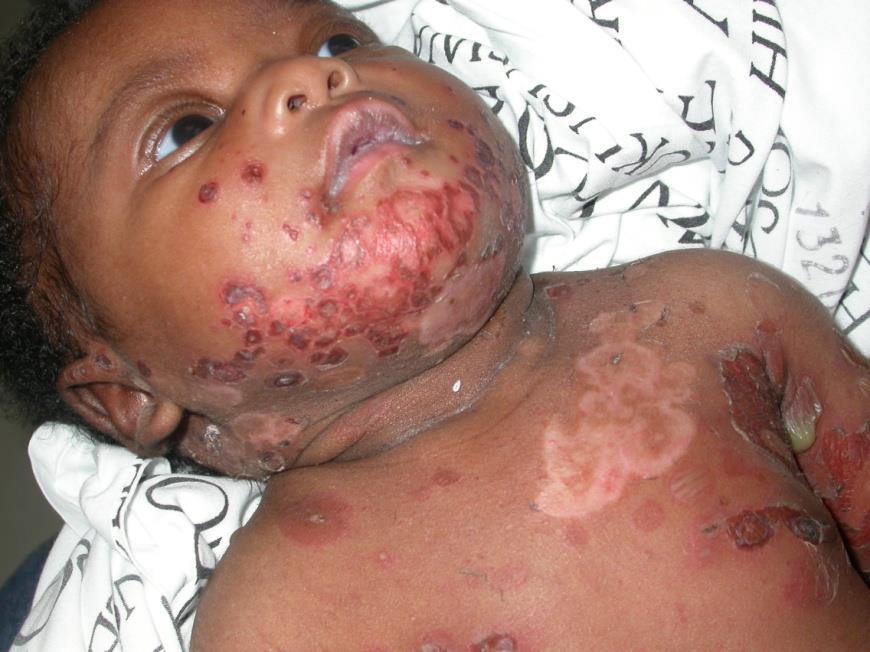
Mimics
- Staphylococcal scalded skin syndrome — Bullous impetigo and SSSS are caused by the same toxins; SSSS is generalised, the child is febrile and miserable, and the skin shears
- Eczema herpeticum — Crusted lesions on eczema may be herpes simplex rather than bacteria - look for monomorphic punched-out erosions and treat urgently if in doubt
- Atopic eczema and infected eczema — Impetiginised eczema is both diagnoses at once; treating only the infection leaves the cause
- Hand, foot and mouth disease — Perioral lesions can look similar early on
What to do in the ED
- Treat per NICE NG153 and local policy. 1.1.2: hydrogen peroxide 1% cream first-line for localised non-bullous impetigo in someone who is not systemically unwell or at high risk of complications. 1.1.3: a short course of a topical antibiotic only if hydrogen peroxide 1% cream is unsuitable. Widespread or bullous disease, or a patient who is systemically unwell or at high risk of complications, takes an antibiotic. 1.1.10: if it worsens or fails to improve on hydrogen peroxide, step to a topical antibiotic while localised, or a topical or oral antibiotic once widespread
- Examine and treat the underlying skin condition, not just the infection - most commonly eczema
- Consider and exclude eczema herpeticum in any child with eczema and crusted lesions; if in real doubt, start aciclovir as well
- Swab if it is recurrent, extensive, or has failed treatment
- Advise on hygiene: separate towels and flannels, hand washing, avoid scratching, cover lesions where practical
- Advise exclusion from school or nursery until all blisters have formed scabs, or 2 days after starting antibiotics (UKHSA wording). CKS words the same rule as 48 hours after commencing treatment, hydrogen peroxide cream or antibiotics - use that, so first-line treatment does not cost the child extra days off
Disposition
Treat and discharge. Admit only for systemic illness, extensive bullous disease in an infant, or when staphylococcal scalded skin syndrome is a possibility.
Safety-netting
Sources
- NICE CKS - Impetigo
- UKHSA - Health protection in children and young people's settings, including education
- Lim HW et al. International Expert Consensus on Knowledge Gaps in Care for Dermatologic Disorders in Skin of Color. Int J Dermatol 2026
- NICE NG153 - Impetigo: antimicrobial prescribing
- NICE CG153 1.2.1.5 - erythema may be underestimated in skin types V-VI (stated in the psoriasis guideline; cited here for that general point about assessing erythema, not for this condition)