Erythema multiforme major
Target lesions on the limbs with mucosal erosions - usually herpes-driven, and not the same disease as SJS.
Red flags
- Extensive mucosal involvement preventing eating, drinking or passing urine
- Ocular involvement - same-day ophthalmology
- Truncal onset, dusky flat macules, detachable epidermis or a positive Nikolsky sign - this is epidermal necrolysis, not EM major, and the destination is different
- A new drug in the preceding weeks with fever and mucosal erosions - again, reconsider epidermal necrolysis
- Systemic upset, dehydration, or respiratory symptoms suggesting mycoplasma
Recognise
Typical targets: a dusky or blistered centre, a paler oedematous middle ring, and a sharply defined red outer ring, raised and palpable, distributed acrally and symmetrically.
The three-zone target that defines the lesion is described entirely in colour, and in brown and black skin the outer ring may be violaceous, hyperpigmented or invisible, so the diagnosis is frequently delayed or missed. Rely on the architecture rather than the colour: run a finger over the lesion, because the concentric zones can be felt as a raised oedematous ring around a flatter, sometimes blistered, centre. The acral symmetrical distribution and the mucosal erosions are tone-independent. Resolution leaves prominent and long-lasting post-inflammatory hyperpigmentation, which is a common reason for re-presentation and is not active disease.
- Raised, palpable target lesions with concentric zones - palpate, do not only look
- Acral, symmetrical distribution spreading inwards, unlike the truncal onset of SJS/TEN
- Mucosal erosions, most often the lips and mouth, define the major form
- A preceding cold sore or herpes simplex infection in most cases, and mycoplasma in children
- Usually a drug is NOT the cause, which is the practical difference from epidermal necrolysis
- In the consensus classification the deciding word is localised versus widespread: bullous erythema multiforme is localised typical or raised atypical targets with under 10% detachment, whereas SJS is widespread purpuric macules or flat atypical targets
Distribution
Photographs
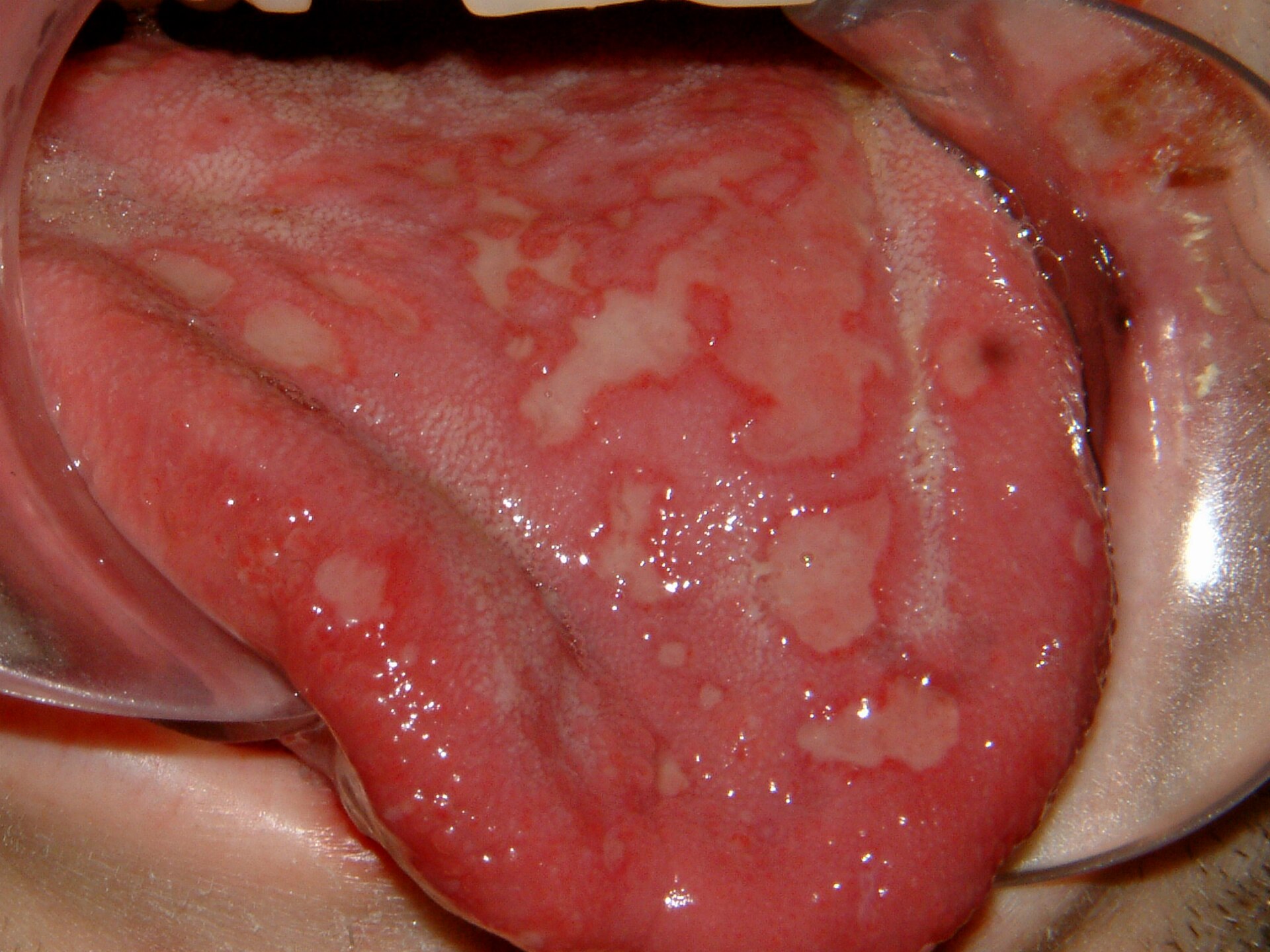
Mimics
- Stevens-Johnson syndrome / toxic epidermal necrolysis — The distinction that matters most on this page. EM major is acral, raised and palpable, usually herpes-driven; SJS/TEN is truncal, flat and dusky, drug-driven, with detachable epidermis - and goes to a burns centre
- Pemphigus vulgaris and bullous pemphigoid - the acute presentation — Both erode mucosa; immunobullous disease evolves over weeks and lacks target lesions
- Eczema herpeticum — Both herpes-associated, but herpeticum gives disseminated monomorphic punched-out erosions on eczematous skin rather than acral targets
What to do in the ED
- Decide explicitly whether this is EM major or epidermal necrolysis, and record the reasoning - distribution, whether lesions are raised, and whether epidermis is detachable
- Look for and treat the trigger: ask about cold sores, and consider mycoplasma in a child with respiratory symptoms
- Assess oral intake and hydration; mucosal pain is what usually determines admission
- Analgesia, mouth care and a topical anaesthetic or barrier as appropriate; ophthalmology the same day for any eye involvement
- Refer to dermatology. Admit if oral intake cannot be maintained, if there is extensive mucosal disease, or if the diagnosis is not secure
- Warn about recurrence, which is common with herpes-associated disease, and refer for consideration of suppressive antiviral treatment if attacks are frequent
Disposition
Urgent dermatology. Admit where mucosal involvement prevents adequate oral intake, where there is ocular involvement, or where epidermal necrolysis has not been confidently excluded - in which case manage as necrolysis until it has.
Safety-netting
Local variation
Sources
- Creamer D et al. U.K. guidelines for the management of Stevens-Johnson syndrome/toxic epidermal necrolysis in adults 2016. Br J Dermatol 2016
- Bastuji-Garin S et al. Clinical classification of cases of toxic epidermal necrolysis, Stevens-Johnson syndrome, and erythema multiforme. Arch Dermatol 1993
- NICE CKS - Herpes simplex - oral
- Lim HW et al. International Expert Consensus on Knowledge Gaps in Care for Dermatologic Disorders in Skin of Color. Int J Dermatol 2026
- NICE CG153 1.2.1.5 - erythema may be underestimated in skin types V-VI (stated in the psoriasis guideline; cited here for that general point about assessing erythema, not for this condition)