Granuloma annulare
A ring of firm papules with no scale - benign, self-limiting and treated as ringworm for months because of the annular shape.
Red flags
- Widespread or generalised disease - worth dermatology assessment rather than reassurance alone, and a firmer prompt to screen for the associated conditions. The cited review reports the comorbidity associations for granuloma annulare as a whole and a greater quality-of-life impact when widespread; it does not grade comorbidity by extent, so no such gradient is claimed here
- Ulceration, pain or rapid growth - this is not granuloma annulare and needs a different pathway
- Lesions on the ear, nose or a chronically sun-exposed site in an older patient that are growing - do not assume a benign annular lesion, and refer
- A patient with the eruption who has not had a recent glucose and lipid check - granuloma annulare is associated with diabetes, hyperlipidaemia and autoimmune disease, so this is a prompt for the GP rather than an ED investigation
Recognise
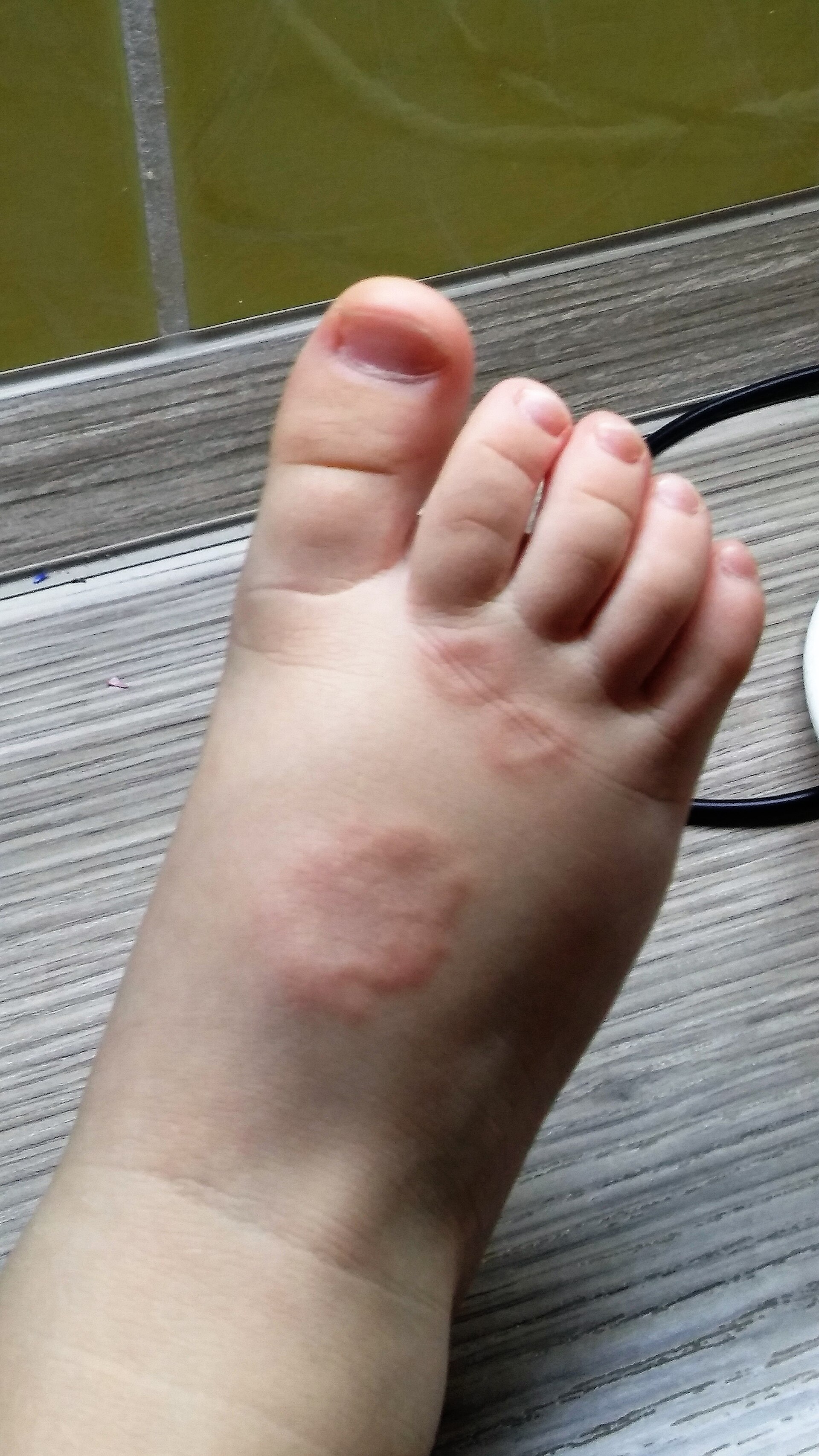
Rings or arcs of small, firm, smooth papules, skin-coloured to pink or red, with a clear centre, on the backs of the hands and feet - and no scale at all.
The papules commonly appear violaceous, dark brown or simply skin-coloured rather than the erythematous ring of the textbook description, so the lesion can read as a patch of pigmentation and the raised structure is easy to miss on inspection. That colour description is clinical observation rather than a cited finding - the review cited here describes granuloma annulare as erythematous, which is itself the problem. Feel the edge: the diagnosis is a palpable rim of firm, smooth papules around a normal centre, and that is entirely tone-independent. The single most useful discriminator is also tone-independent - there is no scale - though that test weakens in anyone who has already applied a topical steroid, because steroid-modified tinea loses its scale and its defined edge. Post-inflammatory hyperpigmentation persists after the lesions flatten and is frequently mistaken for continuing disease.
- An annular plaque with a palpably raised, beaded edge and a normal-looking centre
- No scale, anywhere on the lesion - the discriminator from tinea corporis. But ask what has already been put on it: a lesion treated with a topical steroid may have lost its scale (tinea incognito), so absence of scale means much less in a steroid-exposed lesion
- Not itchy, or only slightly so, and not painful - which also separates it from tinea and from eczema
- Firm and smooth on palpation; the papules feel deeper than they look
- Usually on the backs of the hands and feet and over joints, and often asymmetrical
- Self-limiting in most people, though it may take months to years, and it recurs
Distribution
Photographs
Mimics
- Tinea corporis, cruris and pedis — The mistake that actually happens. Both are annular with a raised edge, but tinea corporis scales at that edge and itches; granuloma annulare has no scale. Months of antifungal for granuloma annulare is a common history
- Lichen planus — Annular lichen planus also gives violaceous papules, but they are flat-topped and shiny with Wickham striae and are usually itchy
- Pityriasis rosea — Both give oval or annular truncal lesions; pityriasis rosea has a trailing collarette of scale and a self-limiting course over weeks
- Lyme disease and erythema migrans — Erythema migrans is a single expanding ring that grows over days after a tick bite, is flat rather than beaded, and needs antibiotics urgently
- Psoriasis, including guttate — Annular psoriasis has thick adherent scale and a well-demarcated flat plaque rather than a beaded papular rim
What to do in the ED
- Run a finger over the edge and look for scale, then ask what has already been applied to it. A raised, beaded, scale-free rim in a steroid-naive lesion is granuloma annulare; a scaling advancing edge is tinea
- Take scrapings for mycology unless the lesion is classical and has had no steroid on it. A negative scrape costs nothing; a tinea missed because a steroid flattened its scale costs months. Do not commit to a long antifungal course on appearance alone either
- Reassure explicitly: this is benign, not infectious, not contagious, and usually self-limiting - though it may take months to years and it can recur
- Do not prescribe an antifungal for it, and stop one that has been started for it and has not worked
- Ask the GP to check glucose and lipids, and to consider thyroid function - the recognised associations are diabetes mellitus, hyperlipidaemia and autoimmune disease including thyroiditis, rheumatoid arthritis and lupus
- Refer to dermatology for widespread disease, diagnostic uncertainty, or where the appearance is causing significant distress - the cited review notes there are no treatments approved by the US Food and Drug Administration, and UK licensing is not stated there, so treatment is a specialist conversation
- Warn about the pigment change that follows, particularly in brown and black skin, so the flattened lesion is not read as active disease
Disposition
Reassure and discharge for typical localised disease, with GP follow-up for the metabolic and autoimmune associations. Routine dermatology referral for widespread disease, diagnostic uncertainty or significant distress.
Safety-netting
Sources
- Foroohar T et al. Granuloma annulare: an updated review of epidemiology, molecular pathogenesis, and management. J Am Acad Dermatol 2026
- Lim HW et al. International Expert Consensus on Knowledge Gaps in Care for Dermatologic Disorders in Skin of Color. Int J Dermatol 2026
- NICE CG153 1.2.1.5 - erythema may be underestimated in skin types V-VI (stated in the psoriasis guideline; cited here for that general point about assessing erythema, not for this condition)