Venous eczema, lipodermatosclerosis and venous leg ulcers
The commonest thing mistaken for bilateral cellulitis - and the single biggest source of unnecessary antibiotics in the ED.
Red flags
- Genuine superimposed cellulitis - unilateral deterioration, increasing pain, fever and systemic upset on a background of chronic venous change
- Absent pulses or an ankle-brachial pressure index that has not been checked before compression - compression bandaging on an arterial leg causes tissue loss
- An ulcer with rolled or everted edges, or one that is not healing as expected - consider malignancy and the need for biopsy
- Sudden unilateral swelling and pain - exclude deep vein thrombosis
- Rapidly progressive pain out of proportion - necrotising infection can arise in a chronically damaged leg
Recognise
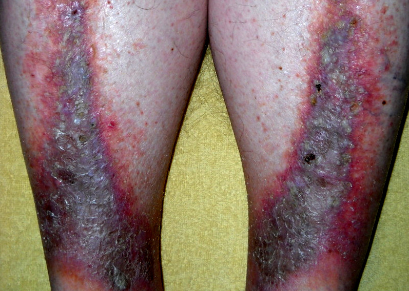
Red-brown discolouration with haemosiderin staining, scaling, itch and oedema over the gaiter area; lipodermatosclerosis produces a hard, tight, indurated 'inverted champagne bottle' leg.
Haemosiderin staining is described as red-brown and is far less distinguishable against brown and black skin, where it merges with the surrounding tone - so the most recognisable feature of the condition disappears. Rely on the structure and the symmetry: bilateral involvement, oedema, the woody induration of lipodermatosclerosis, the inverted champagne bottle shape, varicosities, and scaling with itch rather than pain. Palpate both legs and compare. This matters more here than almost anywhere else in the atlas, because the alternative diagnosis is cellulitis, and a patient with dark skin and bilateral venous disease is at real risk of repeated courses of antibiotics for an infection they do not have. Post-inflammatory hyperpigmentation compounds the confusion further.
- Bilateral - true cellulitis is almost always unilateral, and this is the most useful discriminator in the whole entry
- Itchy rather than painful - cellulitis hurts
- Chronic, with a history over months or years, rather than an acute onset over hours or days
- Oedema, varicosities, haemosiderin change and scaling
- Lipodermatosclerosis: hard, tight, tender, indurated skin with the leg narrowing above the ankle
- Usually afebrile with normal inflammatory markers - but a normal CRP does not exclude cellulitis, particularly early disease, or in an older or immunosuppressed patient
Distribution
Photographs
Mimics
- Cellulitis — The key distinction. Cellulitis is unilateral, acute, painful and often febrile; venous eczema is bilateral, chronic and itchy. Bilateral lower-limb cellulitis is rare
- Contact dermatitis - allergic and irritant — Contact allergy to bandages, dressings or topical antibiotics is very common in this group and perpetuates the eczema
- Necrotising fasciitis and necrotising soft tissue infection — Disproportionate pain on a chronically damaged leg still means escalate
- Atopic eczema and infected eczema — Venous eczema is a distinct pattern driven by venous hypertension, not atopy
- Diabetic foot ulcer and deep-seated foot infection — Venous ulcers sit in the gaiter area and are usually painful; neuropathic ulcers sit on plantar pressure points and are usually painless - and the diabetic foot has a one-working-day referral clock
What to do in the ED
- Ask whether this is actually infection. Bilateral, itchy, chronic and afebrile is usually venous disease rather than cellulitis, and antibiotics will not help that. Then look separately for infection superimposed on one leg - CKS lists infection among the complications of venous disease and asks you to assess for it, so it coexists with the venous pattern rather than being excluded by it
- Emollient generously and a topical corticosteroid for the eczematous component
- Check the arterial supply before anyone applies compression - an ankle-brachial pressure index is needed, and compression on an arterial leg is harmful
- Arrange compression therapy and leg-ulcer service referral where there is an ulcer, since compression is the definitive treatment for venous ulceration
- Elevate the leg and advise on elevation and mobility at home - this treats the underlying cause
- Consider a contact allergy to dressings or topical preparations in eczema that is not settling, and refer for patch testing
- Refer for venous assessment, since treating the underlying venous incompetence reduces recurrence
Disposition
Topical treatment, elevation and referral to community leg-ulcer or tissue viability services. Admit only for genuine superimposed infection, suspected DVT or critical limb ischaemia.
Safety-netting
Local variation
Sources
- NICE CKS - Venous eczema and lipodermatosclerosis
- NICE CKS - Leg ulcer - venous
- NICE NG141 - Cellulitis and erysipelas: antimicrobial prescribing
- Lim HW et al. International Expert Consensus on Knowledge Gaps in Care for Dermatologic Disorders in Skin of Color. Int J Dermatol 2026
- NICE CG153 1.2.1.5 - erythema may be underestimated in skin types V-VI (stated in the psoriasis guideline; cited here for that general point about assessing erythema, not for this condition)