Cellulitis
One of the most over-diagnosed conditions in the ED - and in darker skin, simultaneously one of the most under-diagnosed.
Red flags
- Pain out of proportion to the appearance, rapid progression, crepitus, haemorrhagic bullae or skin anaesthesia - necrotising soft tissue infection, a surgical emergency
- Sepsis, hypotension or a rising lactate
- Periorbital or orbital involvement - a different pathway, needing urgent imaging and ENT or ophthalmology
- Failure to improve on appropriate antibiotics - reconsider the diagnosis before escalating the antibiotic
- Lymphangitis, or a possible uncommon pathogen after a penetrating injury, exposure to water-borne organisms, or an infection acquired outside the UK - both are explicit NG141 1.1.14 referral triggers, and lymphangitis is easy to note and then not act on
- A baby under 1 month - NG141 table 2 gives no antibiotic choice for this age group at all: antibiotic choice is based on specialist advice
- Immunosuppression, diabetes with a foot ulcer, or a prosthetic joint beneath the affected area
Recognise
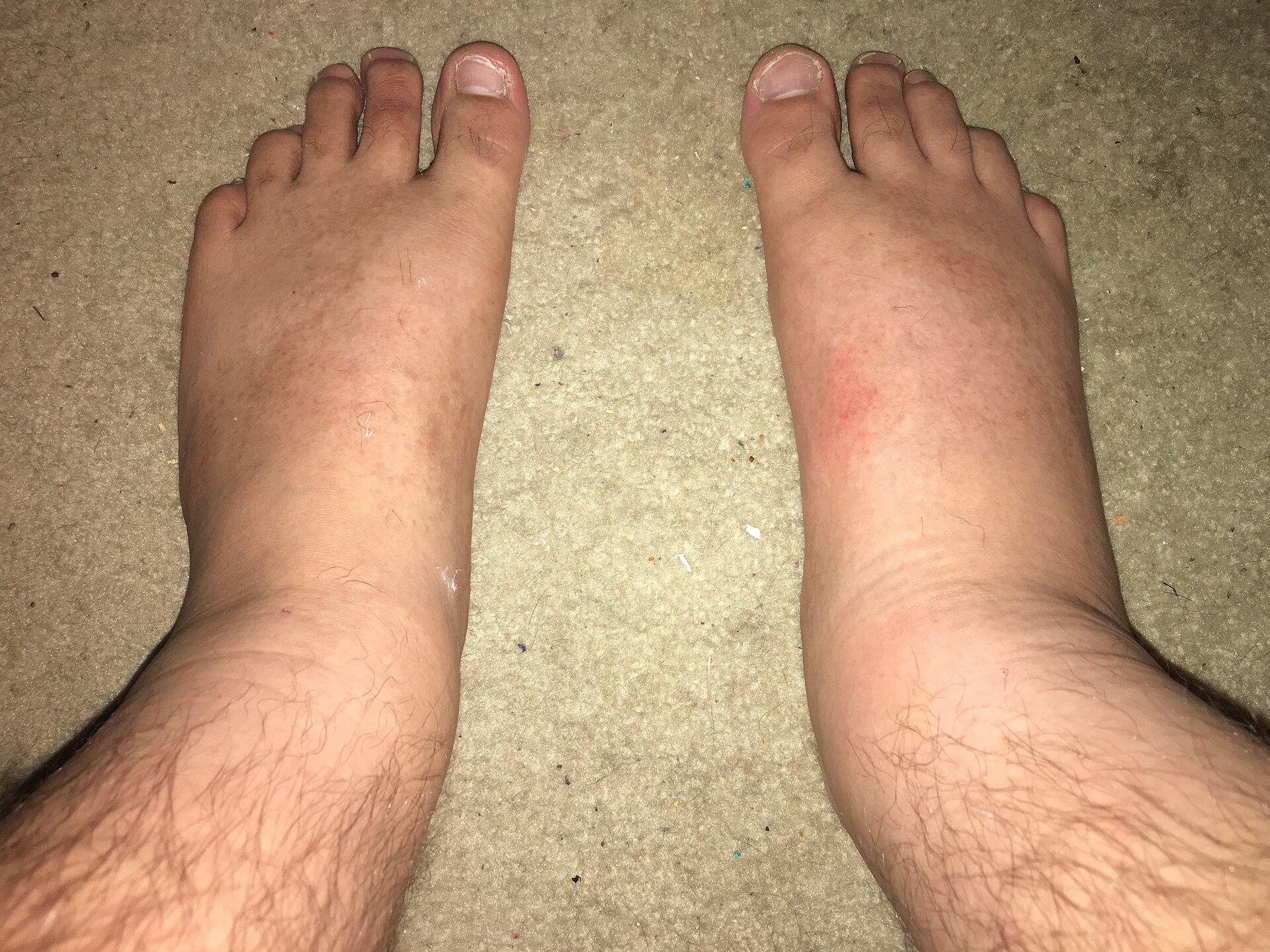
Diffuse warm, tender, spreading erythema with a poorly defined edge, sometimes with oedema, blistering or lymphangitis, in a patient who may be febrile.
Erythema is the entire basis of the textbook description and it is frequently invisible in brown and black skin. Inflamed skin may appear darker, violaceous, greyish or simply shiny rather than red, and this produces error in both directions: cellulitis is missed and treated late, and once suspected it is harder to judge whether it is improving. Use the tone-independent signs - warmth compared with the same site on the other limb, swelling, tenderness and induration - and use the back of your hand to compare temperature side by side. When you mark the margin, use a skin marker and note the anatomical landmark in the notes as well, because a margin drawn on skin whose colour you cannot see is hard for the next clinician to interpret. Photograph with consent where local policy allows, since serial comparison is more reliable than recalled colour.
- Unilateral - bilateral lower-limb cellulitis is rare, and bilateral disease is usually venous eczema or stasis change
- Warmth, swelling and tenderness, with a poorly demarcated advancing edge
- Look for a portal of entry: tinea pedis between the toes, a wound, an ulcer, an insect bite, eczema
- Fever and systemic upset support the diagnosis but are often absent
- Pain out of proportion, rapid progression, crepitus or dusky change means stop and consider necrotising infection
Distribution
Photographs
Mimics
- Necrotising fasciitis and necrotising soft tissue infection — The one that kills. Early necrotising infection looks exactly like cellulitis - disproportionate pain and rapid progression are the discriminators
- Erysipelas — Erysipelas is more superficial, with a sharply demarcated raised edge, and is often facial
- IgA vasculitis (Henoch-Schonlein purpura) — Palpable purpura over the legs is vasculitis, not infection
- Tinea corporis, cruris and pedis — Tinea pedis is frequently the portal of entry and must be treated as well, or the cellulitis recurs
- Diabetic foot ulcer and deep-seated foot infection — A hot swollen foot in a person with diabetes is a diabetic foot problem, not simple cellulitis - and acute Charcot arthropathy mimics infection exactly, often with the skin intact
What to do in the ED
- Ask first whether this is cellulitis at all. Bilateral, chronic, itchy or non-tender presentations are usually venous eczema, lipodermatosclerosis or a deep vein thrombosis, and antibiotics will not help any of them. But that reasoning applies to a well patient only. Fever, tachycardia, a rising lactate or systemic upset overrides the pattern - bilateral cellulitis does occur, venous eczema and stasis change can be superinfected, and NG141 makes systemic illness a referral trigger whatever the distribution. Treat and escalate first, and settle the diagnosis afterwards
- Consider and exclude necrotising infection before settling on cellulitis
- Mark the margin with a skin marker, record the time and describe it against an anatomical landmark rather than by colour alone
- Treat per NICE NG141 and local policy, choosing oral or intravenous route on severity rather than on appearance
- Look for and treat the portal of entry, particularly interdigital tinea pedis - failing to do so is the commonest reason cellulitis recurs
- Elevate the limb and give analgesia; these are part of treatment, not an afterthought
- Arrange review within 48 hours for anyone managed at home, and say explicitly what failure to improve should prompt
Disposition
Oral antibiotics and planned review for uncomplicated disease in a well patient. Admit or refer for intravenous therapy for systemic upset, rapid progression, failure of oral treatment, significant comorbidity, or any suspicion of a deeper infection.
Safety-netting
Local variation
Sources
- NICE NG141 - Cellulitis and erysipelas: antimicrobial prescribing
- NICE CKS - Cellulitis - acute
- Lim HW et al. International Expert Consensus on Knowledge Gaps in Care for Dermatologic Disorders in Skin of Color. Int J Dermatol 2026
- Mukwende M, Tamony P, Turner M. Mind the Gap: A handbook of clinical signs in Black and Brown skin. St George’s, University of London (2020)