Necrotising fasciitis and necrotising soft tissue infection
A surgical emergency masquerading as cellulitis - the diagnosis is clinical suspicion and the treatment is the operating theatre.
Red flags
- Pain out of proportion to the visible skin - escalate on this alone
- Any crepitus, haemorrhagic bullae, skin anaesthesia or dusky change
- Rapid progression, or deterioration despite intravenous antibiotics
- Sepsis with a soft tissue source - treat as necrotising until surgically excluded
Recognise
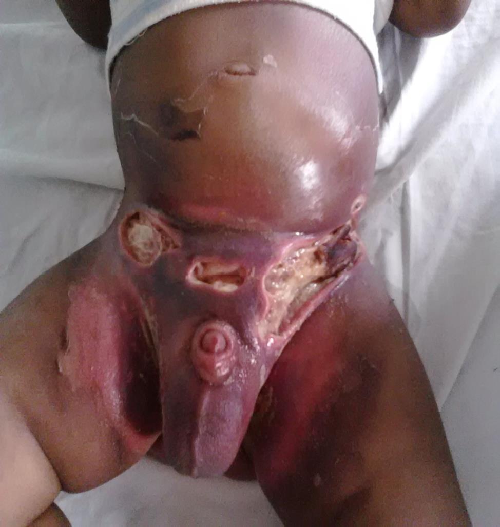
Early on, tense erythema indistinguishable from cellulitis; later a dusky, grey or mottled purple discolouration, haemorrhagic bullae, and skin anaesthesia over the affected area.
The early erythema that prompts the cellulitis diagnosis may be invisible, and the later dusky change is far harder to see against brown or black skin - both of which delay a diagnosis where delay measures directly in mortality. Do not wait for a colour change. Palpate: oedema extending beyond any visible margin, woody induration, crepitus and exquisite tenderness are all tone-independent, as is the pain-out-of-proportion that is the single most useful early sign. Skin anaesthesia over the area is a late but tone-independent finding.
- Pain grossly out of proportion to the visible findings - the earliest and most useful sign
- Rapid progression over hours, with a spreading margin you can mark and watch move
- Systemic toxicity out of keeping with the appearance of the skin
- Oedema or induration extending beyond the visible change
- Crepitus, skin anaesthesia, or haemorrhagic bullae - all late, all mean go to theatre
- Failure to improve on appropriate intravenous antibiotics for cellulitis
Distribution
Photographs
Mimics
- Meningococcal septicaemia and purpura fulminans — Both give purpura with shock; meningococcal purpura is widespread and multifocal rather than centred on one painful compartment
- Toxic shock syndrome — Streptococcal TSS and necrotising fasciitis frequently coexist - finding one is a reason to look for the other
- Cellulitis — The deferred link now closed. Early necrotising infection is indistinguishable from cellulitis on appearance - disproportionate pain and rapid progression are what separate them
- Diabetic foot ulcer and deep-seated foot infection — A diabetic foot with pain out of proportion, crepitus or rapid progression is a surgical emergency first and a diabetic foot pathway second
What to do in the ED
- Escalate to the on-call surgical team immediately on clinical suspicion - do not wait for imaging or bloods
- Resuscitate in parallel and treat as sepsis: fluids, cultures, lactate, and broad-spectrum intravenous antibiotics with anti-anaerobic and anti-toxin cover per local policy
- Mark the margin with the time and re-examine, so progression is measured rather than remembered
- Do not use LRINEC to rule out - see the caveat, this is the most important single point on this page
- CT if it will not delay surgery, recognising that imaging cannot exclude the diagnosis either
- Alert critical care early; definitive treatment is surgical debridement, and delay increases mortality
Score
Disposition
Immediate surgical referral for debridement, with critical care involvement. Admission is to theatre, not to a ward.
Local variation
Sources
- Wong CH et al. The LRINEC (Laboratory Risk Indicator for Necrotizing Fasciitis) score: a tool for distinguishing necrotizing fasciitis from other soft tissue infections. Crit Care Med 2004
- Fernando SM et al. Necrotizing Soft Tissue Infection: Diagnostic Accuracy of Physical Examination, Imaging, and LRINEC Score: A Systematic Review and Meta-Analysis. Ann Surg 2019
- NICE NG141 - Cellulitis and erysipelas: antimicrobial prescribing
- Lim HW et al. International Expert Consensus on Knowledge Gaps in Care for Dermatologic Disorders in Skin of Color. Int J Dermatol 2026
- Barbieri E et al. A population database analysis to describe the residual burden of varicella in Italy, 2004-2022. Front Public Health 2025
- Chiappini E et al. Management of Invasive Group A Streptococcal Infections in Children: ESPID Guidelines. Pediatr Infect Dis J 2026
- NICE CG153 1.2.1.5 - erythema may be underestimated in skin types V-VI (stated in the psoriasis guideline; cited here for that general point about assessing erythema, not for this condition)