Staphylococcal scalded skin syndrome
A toxin-mediated blistering illness of young children that spares the mucosa - the distinguishing feature from epidermal necrolysis.
Red flags
- Mucosal involvement - if the mucosa is involved this is not SSSS; reconsider epidermal necrolysis
- Extensive denudation with fluid loss or temperature instability
- An adult presentation - rare, usually immunosuppressed or in renal failure, and substantially more dangerous
- A neonate - lower threshold for admission and for treating as sepsis
Recognise
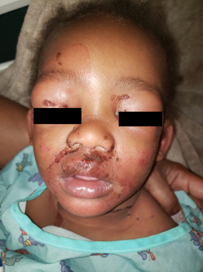
Tender, bright red skin with flaccid blisters that rupture to leave moist denuded areas resembling a scald, with perioral crusting and fissures radiating from the mouth.
The scalded appearance is described in terms of redness that may not be apparent; look instead for the shine and moistness of denuded skin, which is tone-independent, and for the perioral crusting and radial fissuring. Skin tenderness in a miserable, febrile child is the key clinical sign and does not depend on pigment. The denuded areas may look darker or dusky rather than red.
- Exquisite skin tenderness - the child is inconsolable and does not want to be handled
- Perioral crusting with fissures radiating from the mouth
- Flaccid blisters and superficial peeling, with a positive Nikolsky sign
- The mucosa is spared - this is the single most useful discriminator from SJS/TEN
- Flexural onset, fever and irritability
- Most cases are in children under five - conventionally stated, and not a figure from the systematic review cited below, which excluded neonates under 90 days. In adults it is rare and usually associated with renal impairment or immunosuppression, and carries a much higher mortality
Distribution
Photographs
Mimics
- Stevens-Johnson syndrome / toxic epidermal necrolysis — The decisive difference is mucosal involvement: present in necrolysis, absent in SSSS
- Toxic shock syndrome — Both are staphylococcal toxin-mediated; TSS is shocked with a diffuse blanching erythroderma rather than blistering
- Eczema herpeticum — Both give a weeping, crusted, painful eruption in a young child; herpeticum shows monomorphic punched-out erosions, often on eczematous skin
- Erythroderma — Generalised erythema with desquamation in a child may be either; look for the shearing and the perioral crusting
What to do in the ED
- Start intravenous anti-staphylococcal antibiotics per local policy - this is toxin-mediated disease from a staphylococcal focus, which may be small and remote. The 2025 systematic review cited below found clindamycin did not improve outcomes in paediatric SSSS, supporting a beta-lactam as first line - follow local policy, but do not assume clindamycin is needed for anti-toxin cover
- Hunt for the source: umbilicus in neonates, conjunctivae, nose, throat, ears, or any wound
- Swab the likely focus, the nose and throat; blisters themselves are usually sterile, so a negative blister swab means nothing
- Analgesia before handling, and handle as little as possible - this is a genuinely painful condition and is often undertreated
- Fluid balance, temperature control, and non-adherent dressings to denuded areas
- Admit under paediatrics, with dermatology input; involve burns for extensive denudation
- Check the mucosa explicitly and document that you did, because that examination is what separates this from a burns-centre diagnosis
Disposition
Admit under paediatrics for intravenous antibiotics, analgesia and fluid balance. Burns input for extensive skin loss. Adults need admission and a search for underlying immunosuppression or renal failure. Scope note on the evidence here: the paediatric systematic review cited below deliberately excluded neonates under 90 days and preterm infants, so the neonatal content on this page comes from the separate neonatal literature and the adult content from a separate adult-versus-paediatric comparison. Adult disease is widely described as carrying a higher mortality than childhood disease; that comparison is conventional and we have not been able to source a figure for it, so it is given here as a reason to admit rather than as a number.
Sources
- Gray L et al. Pediatric Staphylococcal Scalded Skin Syndrome: A Systematic Review of the Literature to Inform Work-Up and Management. Pediatr Dermatol 2025 (excluded neonates under 90 days and preterm infants)
- Jovic TH et al. Desquamating dermatological conditions: The burns surgeon's guide to diagnosis and management. J Plast Reconstr Aesthet Surg 2026
- Lim HW et al. International Expert Consensus on Knowledge Gaps in Care for Dermatologic Disorders in Skin of Color. Int J Dermatol 2026
- Lorenzin G et al. Staphylococcal Scalded Skin Syndrome in a Preterm Infant. Antibiotics (Basel) 2025 - neonatal and preterm susceptibility
- Patel S, Cadwell JB, Lambert WC. Comparison of adult vs. paediatric inpatients with staphylococcal scalded skin syndrome. Br J Dermatol 2021
- NICE CG153 1.2.1.5 - erythema may be underestimated in skin types V-VI (stated in the psoriasis guideline; cited here for that general point about assessing erythema, not for this condition)