Stevens-Johnson syndrome / toxic epidermal necrolysis
Drug-induced epidermal necrolysis with mucosal involvement - a dermatological emergency that belongs in a burns or specialist centre.
Red flags
- Any new blistering or mucosal erosion in a patient on a new drug - treat as epidermal necrolysis until proven otherwise
- Detachable epidermis or a positive Nikolsky sign
- Ocular involvement - but the referral is not conditional on finding it: the UK guideline makes an ophthalmologist part of the initial assessment of every patient, and ocular inflammation may develop before the skin signs
- Rapidly increasing area of detachment between examinations
Recognise
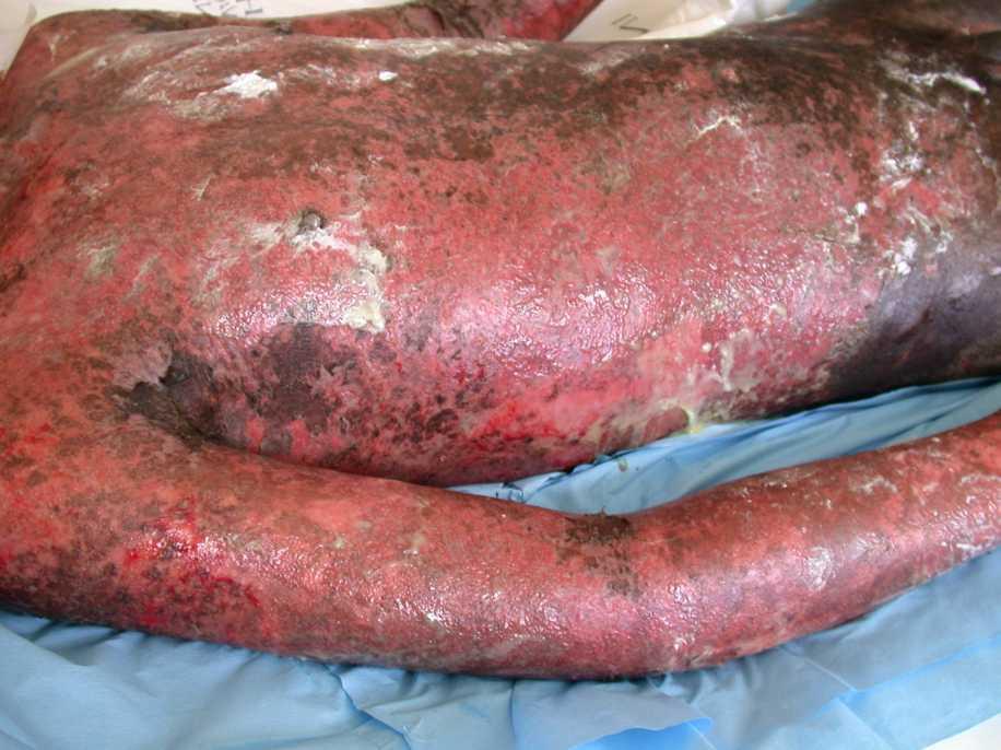
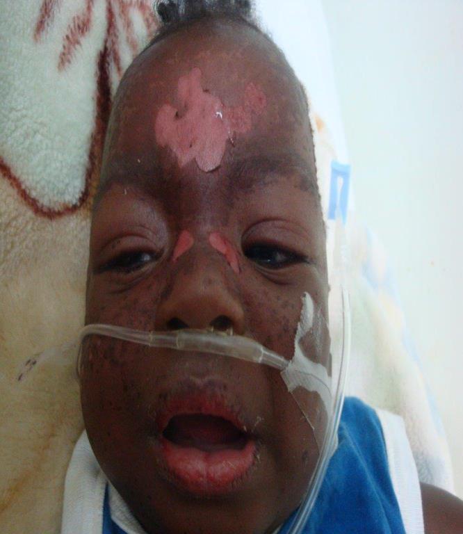
Dusky red or purpuric irregular macules that coalesce, often with a darker centre, progressing to flaccid blisters and sheets of epidermal detachment leaving raw, weeping dermis.
The erythema that defines the early rash in pale skin may be absent, subtle or appear violaceous, grey or hyperpigmented rather than red, so the eruption can look deceptively unimpressive at the stage when it is most treatable. Rely on the tone-independent features instead: severe skin pain out of proportion to what is visible, mucosal erosions, and detachable epidermis. Look for early change on the palms, soles and conjunctivae where pigment is less of an obstacle.
- Skin pain and tenderness out of proportion to the visible rash
- Erosions or crusting of two or more mucosal surfaces - eyes, mouth, genitals
- Positive Nikolsky sign: gentle lateral pressure shears the epidermis
- Prodromal fever and malaise for a few days before the rash
- A new drug started within the preceding weeks, and a culprit is demonstrable in roughly 85% of cases. The high-risk drugs are a short, memorable list: allopurinol, carbamazepine, lamotrigine, nevirapine, oxicam NSAIDs, phenobarbital, phenytoin, sulfamethoxazole and other sulfonamide antibiotics, and sulfasalazine. Risk is concentrated in the first few weeks of exposure
- Do not blame the wrong drug. Paracetamol, aspirin, ibuprofen and corticosteroids have an unclear association and are likely to be confounders - they are the drugs given to treat the prodrome
Distribution
Photographs
Mimics
- Staphylococcal scalded skin syndrome — Both blister and shear. SSSS spares the mucosa and affects young children; SJS/TEN involves mucosa
- Erythema multiforme major — Both have mucosal erosions, but EM major has typical raised target lesions on the limbs and is usually herpes-driven, not drug-driven
- Pemphigus vulgaris and bullous pemphigoid - the acute presentation — Also blisters with a positive Nikolsky in pemphigus, but evolves over weeks rather than days and lacks the drug prodrome
- Acute generalised exanthematous pustulosis — Also an acute drug reaction with fever, but pustules on oedematous erythema rather than detachment
What to do in the ED
- Stop every non-essential drug started in the preceding weeks and document the exact start dates - culprit identification is the single most important intervention
- Map and record the percentage of detached plus detachable epidermis, and repeat it, because the figure drives both prognosis and destination. Record erythema and detachment as two separate numbers on a Lund and Browder chart - scoring erythema as detachment inflates SCORTEN, and it is detachment, not erythema, that carries the prognostic weight
- Calculate SCORTEN within the first 24 hours of admission, from the admission observations and bloods, and recalculate as the picture evolves - an early score is not the final prognosis. The guideline pairs this with drug causality assessment inside the same 24 hours
- Discuss with the regional burns centre and with dermatology now, not after investigations
- Involve an ophthalmologist as part of the initial assessment, within 24 hours, whether or not you can see eye involvement - the UK guideline asks for this in every case, and for daily ocular review through the acute illness. Eye involvement occurred in 74% of patients in the series it cites, and can precede the rash
- Handle the skin as a burn, and stop the things triage does automatically: no sphygmomanometer cuffs, no adhesive ECG leads, no adhesive dressings, no identification wrist tags. Non-adherent dressings, minimal manipulation, barrier nursing in a side room with controlled humidity on a pressure-relieving mattress, and ambient temperature raised to 25-28 C
- Fluid resuscitation guided by losses, analgesia, and mouth care; take cultures but do not give prophylactic antibiotics
- Escalate to critical care if there is airway, ocular or respiratory involvement or haemodynamic instability
Score
Disposition
Discuss with the regional burns centre and dermatology. Above 10% body surface epidermal loss the UK guideline asks for admission without delay to a Burn Centre, or to an intensive care unit experienced in SJS/TEN with the facilities for extensive skin-loss wound care - co-ordinated by a specialist in skin failure, usually dermatology or plastic surgery, with intensive care, ophthalmology and skin-care nursing. Consider later transfer to a Burn Centre in TEN above 30% with clinical deterioration, extending detachment, sub-epidermal pus, local sepsis, wound conversion or delayed healing. Children and young people should be discussed with a paediatric burns or specialist centre. Do not admit to a general ward while awaiting that conversation.
Local variation
Sources
- Creamer D et al. U.K. guidelines for the management of Stevens-Johnson syndrome/toxic epidermal necrolysis in adults 2016. Br J Dermatol 2016
- McPherson T et al. British Association of Dermatologists' guidelines for the management of Stevens-Johnson syndrome/toxic epidermal necrolysis in children and young people, 2018. Br J Dermatol 2019
- Bastuji-Garin S et al. SCORTEN: a severity-of-illness score for toxic epidermal necrolysis. J Invest Dermatol 2000
- Bastuji-Garin S et al. Clinical classification of cases of toxic epidermal necrolysis, Stevens-Johnson syndrome, and erythema multiforme. Arch Dermatol 1993
- Lim HW et al. International Expert Consensus on Knowledge Gaps in Care for Dermatologic Disorders in Skin of Color. Int J Dermatol 2026
- Mockenhaupt M et al. Stevens-Johnson syndrome and toxic epidermal necrolysis: assessment of medication risks with emphasis on recently marketed drugs. The EuroSCAR-study. J Invest Dermatol 2008
- NICE CG153 1.2.1.5 - erythema may be underestimated in skin types V-VI (stated in the psoriasis guideline; cited here for that general point about assessing erythema, not for this condition)