Acute generalised exanthematous pustulosis
An abrupt drug-induced pustular eruption with fever and neutrophilia; looks alarming, usually settles once the drug stops.
Red flags
- Mucosal erosion or detachable epidermis - reconsider epidermal necrolysis, which is a different emergency
- Hypotension or organ dysfunction - look for an alternative or additional diagnosis
- Deranged liver or renal function suggesting overlap with DRESS
Recognise
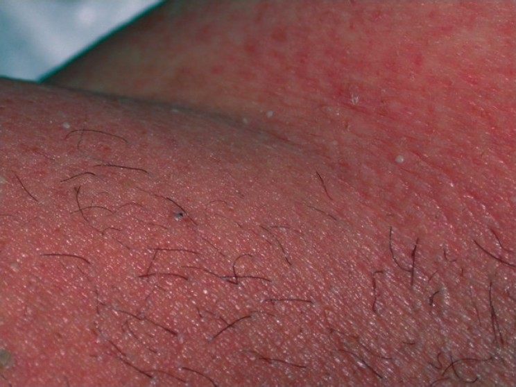
Sheets of small non-follicular sterile pustules studding bright oedematous erythema, beginning in the flexures, followed by desquamation as it resolves.
The pustules themselves are the tone-independent finding and remain visible as pale or yellow-white pinpoints, but the oedematous erythema underneath may not read as red at all - it can look darker, violaceous or simply swollen and shiny, so the classic description of pustules on a red base can be misleading. Feel for the oedema. Resolution leaves marked post-inflammatory hyperpigmentation with the desquamation.
- Onset within days of starting a drug - typically faster than DRESS
- Dozens or more small non-follicular pustules, sterile on culture
- Fever and neutrophilia
- Flexural onset
- Resolves with desquamation over one to two weeks after the drug stops - a conventional interval, not one stated by the EuroSCAR study cited below
Distribution
Photographs
Mimics
- Stevens-Johnson syndrome / toxic epidermal necrolysis — Both acute drug reactions with fever; necrolysis detaches epidermis and erodes mucosa rather than forming pustules
- Generalised pustular psoriasis — The deferred link now closed. Generalised pustular psoriasis presents almost identically; a personal or family history of psoriasis does NOT distinguish them, because the EuroSCAR study found no increased AGEP risk with a psoriasis history. The drug timeline is the discriminator
- Erythroderma — Either condition can become erythrodermic
What to do in the ED
- Stop the likely culprit - antibiotics dominate the list, with ampicillin and amoxicillin, quinolones, anti-infective sulphonamides, terbinafine, (hydroxy)chloroquine and diltiazem the drugs most strongly associated in the EuroSCAR case-control study. Its top-ranked drug, pristinamycin, is omitted here only because it is not marketed in the UK
- Swab or aspirate a pustule: sterility supports the diagnosis and excludes bacterial pustulosis
- Take FBC, liver and renal function - to confirm neutrophilia and to look for overlap with DRESS
- Do not reach for systemic antibiotics for the pustules themselves; they are sterile and an antibiotic may be the cause
- Discuss with dermatology. Admit if febrile and unwell, extensively involved, or if the diagnosis is not secure
- Document the drug as a reaction and avoid re-exposure
Disposition
Same-day dermatology. Admit if febrile, systemically unwell, extensively involved, if liver or renal function is deranged, or if epidermal necrolysis has not been confidently excluded - which, given that fever and neutrophilia are the rule in AGEP rather than the exception, will be most patients at first presentation.
Safety-netting
Sources
- Sidoroff A et al. Risk factors for acute generalized exanthematous pustulosis (AGEP) - results of a multinational case-control study (EuroSCAR). Br J Dermatol 2007
- Lim HW et al. International Expert Consensus on Knowledge Gaps in Care for Dermatologic Disorders in Skin of Color. Int J Dermatol 2026
- NICE CG153 1.2.1.5 - erythema may be underestimated in skin types V-VI (stated in the psoriasis guideline; cited here for that general point about assessing erythema, not for this condition)