Pemphigus vulgaris and bullous pemphigoid - the acute presentation
Two autoimmune blistering diseases that differ in depth, and that difference decides how sick the patient is.
Red flags
- Extensive erosions with fluid loss, pain or secondary infection - barrier failure needs admission
- Inability to eat or drink because of mouth erosions
- Any suggestion this is instead an acute drug reaction - a rapid onset with fever and a new drug points to epidermal necrolysis, which goes to a burns centre
- Sepsis through eroded skin
- Ocular or laryngeal mucosal involvement - suggests mucous membrane pemphigoid and needs urgent specialist assessment
Recognise
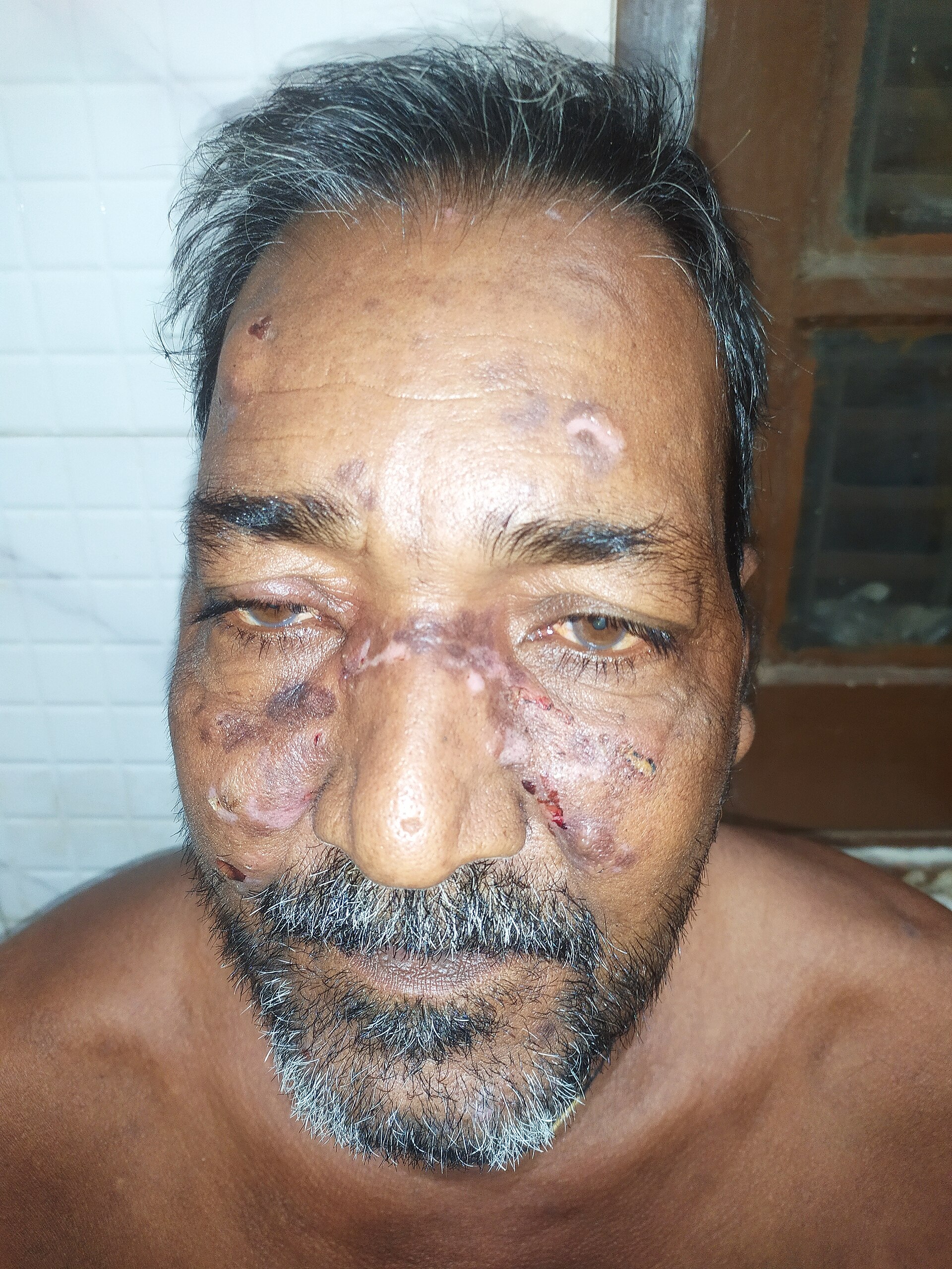
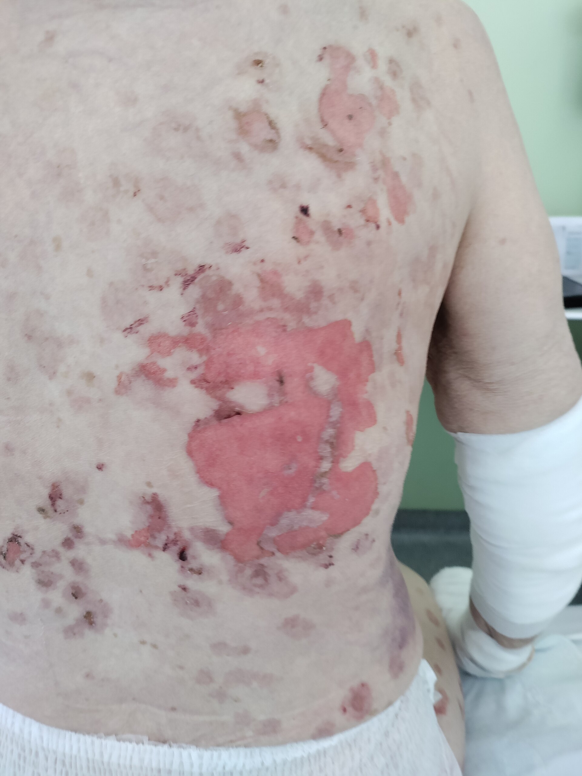
Pemphigus vulgaris: flaccid, fragile blisters that rupture almost immediately, leaving painful raw erosions, with a positive Nikolsky sign. Bullous pemphigoid: tense, dome-shaped blisters on red or urticarial plaques that remain intact for days, intensely itchy, with a negative Nikolsky sign.
The blisters and erosions are tone-independent, but the inflammatory background of bullous pemphigoid is not - the urticarial plaques on which the blisters sit may be violaceous, hyperpigmented or barely visible rather than red, so an early pre-bullous pemphigoid presenting as itch alone is easily dismissed. In pemphigus, mucosal erosions are the presenting feature in most patients and the mouth is unaffected by skin pigment, so examine it. Healing in both leaves conspicuous and long-lasting post-inflammatory hyperpigmentation, which patients may find as troubling as the blisters and which should not be read as active disease.
- Flaccid and fragile, breaking early, mouth involved, Nikolsky positive - pemphigus vulgaris, a more unwell patient with a deeper split
- Tense and intact, very itchy, mouth spared, Nikolsky negative - bullous pemphigoid, usually elderly
- Painful mouth erosions preventing eating and drinking are the commonest first presentation of pemphigus vulgaris
- Bullous pemphigoid may present with weeks of intense itch and urticarial plaques before any blister appears
- Course over weeks, without the acute drug prodrome of epidermal necrolysis
Distribution
Photographs
Mimics
- Stevens-Johnson syndrome / toxic epidermal necrolysis — Both blister with mucosal erosions and a positive Nikolsky in pemphigus; necrolysis is drug-triggered and evolves over days, immunobullous disease over weeks
- Staphylococcal scalded skin syndrome — Both shear superficially with a positive Nikolsky; SSSS is a febrile illness of young children and spares mucosa
- Eczema herpeticum — Both give crusted erosions; herpeticum's erosions are monomorphic, punched-out and on eczematous skin
- Erythema multiforme major — Both give mucosal erosions with blistering; EM major has target lesions and is usually herpes-driven
What to do in the ED
- Establish the pattern before treating: is the blister flaccid or tense, is the mouth involved, is Nikolsky positive - and document it, because the answer determines urgency
- Do not start systemic corticosteroid in the ED without dermatology discussion; treatment is guided by diagnosis, and a skin biopsy with immunofluorescence is needed to confirm it
- Refer to dermatology. Pemphigus vulgaris needs urgent specialist assessment; bullous pemphigoid needs prompt assessment but is less often an emergency
- Dress eroded skin with non-adherent dressings, provide adequate analgesia, and swab if there is any sign of secondary infection
- Mouth care and dietetic input where oral erosions are limiting intake; assess hydration
- Admit for extensive disease, barrier failure, inability to maintain oral intake, or secondary infection
- Take a careful drug history anyway - both conditions have recognised drug-induced forms
Disposition
Urgent dermatology for diagnosis by biopsy and immunofluorescence. Admit for extensive erosion, uncontrolled pain, inability to eat or drink, or secondary infection - pemphigus vulgaris substantially more often than bullous pemphigoid.
Safety-netting
Sources
- Harman KE et al. British Association of Dermatologists' guidelines for the management of pemphigus vulgaris 2017. Br J Dermatol 2017
- Venning VA et al. British Association of Dermatologists' guidelines for the management of bullous pemphigoid 2012. Br J Dermatol 2012
- Lim HW et al. International Expert Consensus on Knowledge Gaps in Care for Dermatologic Disorders in Skin of Color. Int J Dermatol 2026
- NICE CG153 1.2.1.5 - erythema may be underestimated in skin types V-VI (stated in the psoriasis guideline; cited here for that general point about assessing erythema, not for this condition)