Polymorphic light eruption
An itchy papular eruption on newly sun-exposed skin, commoner in brown and black skin than the textbooks imply, and with a variant that is almost peculiar to it.
Red flags
- A photosensitive eruption with systemic features, a malar distribution, mouth ulcers, joint pain or hair loss - consider lupus erythematosus and investigate rather than labelling it sun allergy
- A new photosensitive eruption after starting a drug - a phototoxic or photoallergic drug reaction is a different diagnosis with a different action
- Severe, blistering or scarring photosensitivity, or onset in early childhood - refer, because the inherited photodermatoses and porphyrias present this way
- An eruption on chronically exposed skin including the face and hands, in an older patient, that does not settle - chronic actinic dermatitis, which was also commoner in dark-skinned patients in the same series
Recognise
Itchy pink or red papules, papulovesicles or plaques appearing hours to a couple of days after sun exposure, on skin that has been covered over winter, settling over days if the sun is avoided.
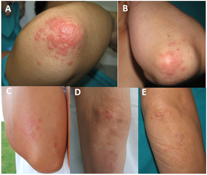
This is a condition the textbooks under-attribute to brown and black skin, and the evidence points the other way. In a 23-year single-centre series of 844 patients, polymorphic light eruption was significantly commoner in those with dark skin types than light (74.0% versus 25.9%) - a clinic-based comparison rather than a population prevalence - and just over half that clinic’s general population was White, which is what makes the imbalance striking - and the same centre found the pattern for chronic actinic dermatitis. The pinpoint papular variant was predominantly seen in dark-skinned individuals, and a separate review describes that variant as peculiar to darker skin. It presents as innumerable tiny monomorphic papules rather than the plaques of the classic description, the erythema is usually not visible, and it is characteristically mistaken for lichen nitidus. Feel the skin: the eruption is palpably rough and papular where it may be nearly invisible. Post-inflammatory hyperpigmentation afterwards is prominent and outlasts the eruption.
- The timing is the diagnosis: hours to two days after sun exposure, on skin newly uncovered - the first hot weekend, a holiday, the start of spring
- Sparing of the face and the backs of the hands is the classic and useful clue, because those sites are exposed all year and have hardened
- Itch is prominent, and the eruption is monomorphic within a given patient even though it is polymorphic between patients
- Settles over days to a couple of weeks if further sun is avoided, without scarring
- The pinpoint papular variant - innumerable tiny papules - is reported predominantly in darker skin and is easily mistaken for lichen nitidus
- Recurs each spring or on each holiday, and often improves through the summer as the skin hardens
- Not the same as prickly heat, though the two are routinely conflated - miliaria arises in covered, occluded, sweaty skin rather than in newly sun-exposed skin, and the distribution separates them
Distribution
Photographs
Mimics
- Lichen planus — The pinpoint papular variant is characteristically mistaken for lichen nitidus, a close relative of lichen planus - the sun-exposed distribution and the seasonal timing are what separate them
- Contact dermatitis - allergic and irritant — A photoallergic contact reaction to sunscreen or a fragrance sits in the same distribution - ask what was applied before the sun exposure
- Drug eruptions - morbilliform and fixed — Phototoxic drug reactions - doxycycline among the commonest - are exaggerated sunburn in a sun-exposed distribution rather than itchy papules
- Acute urticaria — Solar urticaria appears within minutes of exposure and each weal resolves within an hour, rather than appearing after hours and lasting days
What to do in the ED
- Take the exposure history precisely: how long after the sun, what was uncovered, and whether the face and hands are spared. That sequence makes the diagnosis and nothing else is needed
- Examine by palpation in brown and black skin, and look specifically for the pinpoint papular pattern rather than the plaques of the textbook description
- Treat symptomatically - emollient, a topical corticosteroid appropriate to the site for a short course, and an oral antihistamine for itch, recognising that antihistamines help the itch rather than the eruption
- Give the prevention advice, because that is the treatment that matters: high-factor broad-spectrum sunscreen applied properly and reapplied, clothing cover, and graded re-exposure rather than a sudden first long exposure
- Say explicitly that this is not an allergy to sunlight in the anaphylactic sense and that it is not dangerous - patients are frequently frightened by the phrase sun allergy
- Warn about post-inflammatory pigment change before the patient discovers it, particularly in brown and black skin
- Refer to dermatology for severe, disabling or atypical disease, or where prophylactic phototherapy before summer is being considered
Disposition
Reassure, treat symptomatically and discharge. Dermatology referral for severe or atypical disease, where the diagnosis is uncertain, or where a photodermatosis other than polymorphic light eruption is suspected.
Safety-netting
Sources
- Maghfour J, Mohney L, Lim HW, Mohammad TF. Demographics and clinical presentations of 844 patients with light and dark skin types with polymorphous light eruption and chronic actinic dermatitis evaluated over 23 years. Photodermatol Photoimmunol Photomed 2023
- Sharma VK, Sahni K. Photodermatoses in the Pigmented Skin. Adv Exp Med Biol 2017
- Lim HW et al. International Expert Consensus on Knowledge Gaps in Care for Dermatologic Disorders in Skin of Color. Int J Dermatol 2026