Benign neonatal rashes - the reassurance set
Four common, harmless newborn rashes - and the discipline of proving that the baby is well before calling any rash benign.
Red flags
- Any unwell, febrile, poorly feeding or jaundiced neonate - a rash is not the diagnosis, sepsis is, until excluded
- Vesicles, especially clustered or on the scalp or presenting part - neonatal herpes simplex, which is a time-critical emergency and can occur without maternal history
- A rash present with temperature instability, lethargy, or abnormal observations
- Petechiae or purpura in a neonate - never benign without investigation
- Blistering, skin fragility or erosions - consider staphylococcal scalded skin syndrome and congenital blistering disorders
Recognise
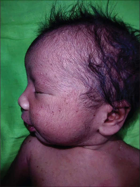
Erythema toxicum shows blotchy red macules with a central white or yellow papule or pustule, appearing in the first few days and migrating. Milia are tiny firm white papules. Transient neonatal pustular melanosis shows superficial pustules that rupture to leave a collarette of scale and a pigmented macule.
The blotchy erythema of erythema toxicum is often invisible in a baby with brown or black skin, leaving only the small pale central pustules on unremarkable skin - so the classic 'flea-bitten' description does not transfer. Transient neonatal pustular melanosis is substantially more frequent in Black infants - Laude 1995 lists it among the disorders commoner in Black children, and a 2012 Dutch report puts it at 0.2 to 2.2% of white neonates against 4 to 5% of black neonates (figures from that paper's abstract; it is a case report, so treat the numbers as indicative of the size of the difference rather than as a UK prevalence) and is frequently the correct diagnosis where it is not considered: pustules present from birth, no surrounding erythema, rupturing to leave a collarette of fine scale and a lasting hyperpigmented macule. Those residual brown macules persist for weeks to months and are routinely mistaken for bruising or for ongoing infection, which causes avoidable investigation and avoidable parental alarm. Dermal melanocytosis is the other finding this entry exists to name. Mind the Gap describes it as patches darker than the surrounding skin, present in newborns and generally gone by age 4, which may look like bruises but are more homogeneous and uniform in colour than a bruise - and it is explicit that history and thorough inspection are needed to avoid misdiagnosing it as non-accidental bruising, and equally to avoid dismissing a real injury as a birthmark. Document the site, size and colour at first presentation so the next clinician is not starting from nothing. Naming both conditions correctly is the whole value of this entry.
- The baby is well, feeding normally, afebrile and has normal observations - this is the precondition for any benign label
- Erythema toxicum: appears day 1 to 3, lesions migrate over hours, baby entirely well
- Transient neonatal pustular melanosis: pustules often present at birth, leaving pigmented macules with a scale collarette
- Milia: tiny firm white papules on the nose and cheeks, resolve spontaneously
- Miliaria: tiny clear or pustular lesions from heat and overwrapping
- Dermal melanocytosis (slate-grey naevi, blue-grey spots, historically Mongolian blue spots): blue-grey or slate-grey patches, most often over the sacrum, buttocks and back, present from birth and usually gone by about age 4. They are normal, and they are the classic benign finding mistaken for bruising and escalated as non-accidental injury - and the mistake runs both ways
- None of these are tender, and none make a baby unwell
Distribution
Photographs
Mimics
- Staphylococcal scalded skin syndrome — Blistering and skin fragility in a neonate is never benign; SSSS in the newborn needs admission and intravenous antibiotics
- Eczema herpeticum — Clustered vesicles in a neonate raise neonatal herpes simplex, which is an emergency
- Impetigo — Bullous impetigo occurs in neonates and can be mistaken for a benign pustular rash
- Nappy rash, candida, and patterns that should worry you — Nappy-area rashes in the newborn have their own differential
What to do in the ED
- Assess the baby, not the rash: temperature, feeding, tone, observations and a full examination
- If the baby is at all unwell, treat as neonatal sepsis and do not attribute anything to a benign rash
- Look specifically for vesicles and ask about maternal genital herpes - but remember that most neonatal herpes occurs with no maternal history, so the absence of one excludes nothing
- In a well baby with a typical benign rash, no investigation is needed
- Name the diagnosis explicitly to the parents, and explain the expected course - particularly the persistent pigmented macules of transient neonatal pustular melanosis, which otherwise generate repeat attendances
- Have a very low threshold for paediatric review in any neonate you are not certain about
Disposition
Discharge only a well, afebrile, normally feeding baby with a clearly typical benign rash and normal observations. Anything else is a neonatal paediatric assessment.
Safety-netting
Sources
- NICE NG143 - Fever in under 5s: assessment and initial management (applies to children under 5 only)
- NICE CKS - Nappy rash
- Laude TA. Approach to dermatologic disorders in black children. Semin Dermatol 1995
- Mukwende M, Tamony P, Turner M. Mind the Gap: A handbook of clinical signs in Black and Brown skin. St George’s, University of London (2020)
- van Delft R, Rosias PR. A neonate with vesicopustular rash [in Dutch]. Ned Tijdschr Geneeskd 2012;156(6):A2816
- Snyder KAM, Voelckers AD. Newborn Skin: Part I. Common Rashes and Skin Changes. Am Fam Physician 2024;109(3):212-216