Molluscum contagiosum
Harmless, self-limiting and enormously common - the intervention is explanation, not treatment.
Red flags
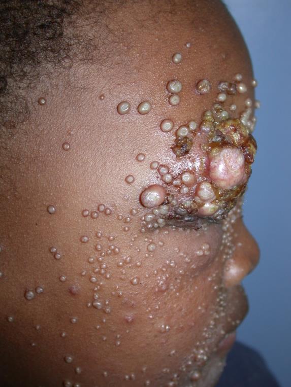
- Very extensive or atypical lesions, or lesions on the face in an adult - consider immunosuppression including HIV
- Genital lesions in a child - consider the possibility of sexual contact and follow safeguarding guidance, while recognising that autoinoculation from elsewhere is common and far more likely
- Lesions near the eye with conjunctivitis
- Genuine secondary bacterial infection with spreading cellulitis
Recognise
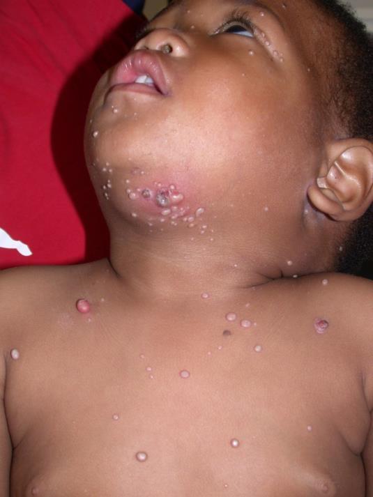
Firm, dome-shaped, pearly pink papules 2 to 5 mm across with a central dimple, sometimes with surrounding eczema.
The pearly pink colour does not transfer; lesions in brown and black skin more often appear skin-coloured, brown or hyperpigmented, which can make them look like warts or other papules. The dome shape and the central umbilication are structural, not chromatic - look across the skin at an angle, in good light, to catch the dimple. Molluscum characteristically leaves conspicuous post-inflammatory hyperpigmentation at each site as lesions resolve, often for months, and parents should be warned that the marks outlast the lesions and do not mean the infection is persisting.
- Central umbilication - the dimple is the diagnosis
- Firm, dome-shaped, usually 2 to 5 mm
- In clusters, spread by direct contact and autoinoculation
- Surrounding eczema is common and is often what brings them in
- An inflamed, red, tender lesion usually means it is about to resolve, not that it is infected
Distribution
Photographs
Mimics
- Chickenpox (varicella) — Chickenpox lesions are vesicular, at mixed stages, and accompanied by fever
- Atopic eczema and infected eczema — Molluscum eczema around lesions is common and both may need treating
- Impetigo — An inflamed molluscum can be mistaken for impetigo and treated with antibiotics unnecessarily
What to do in the ED
- Explain that it is harmless and self-limiting, but be honest about the timescale: CKS states that resolution usually occurs within 18 months, though lesions can persist for more than 3 or 4 years. Understating this is why families lose confidence and re-attend
- Treatment is not routinely recommended - most interventions risk scarring and the condition resolves without them
- Treat surrounding eczema, which is usually the actual complaint
- No exclusion is required - molluscum does not appear in UKHSA's A to Z of infectious diseases in education and childcare settings at all, and where no exclusion period is set the guidance's position is that the person attends and interacts as usual. Swimming need not be restricted either, though that part is conventional advice rather than a UKHSA statement. Advise against sharing towels and against scratching
- Warn that lesions often become red and inflamed shortly before they resolve, and that this is expected rather than infection
- Consider immunosuppression in extensive or atypical disease; assess genital lesions in children within safeguarding guidance without assuming abuse
Disposition
Reassure and discharge. Refer only for extensive or atypical disease, suspected immunosuppression, or where a safeguarding concern arises.
Safety-netting
Sources
- NICE CKS - Molluscum contagiosum
- UKHSA - Health protection in children and young people's settings, including education
- Lim HW et al. International Expert Consensus on Knowledge Gaps in Care for Dermatologic Disorders in Skin of Color. Int J Dermatol 2026
- Mukwende M, Tamony P, Turner M. Mind the Gap: A handbook of clinical signs in Black and Brown skin. St George’s, University of London (2020)