Infantile haemangioma
The commonest benign tumour of infancy, in up to 5% of infants - mostly harmless, but a minority need referral within weeks and the window to act is short.
Red flags
- Periorbital or periocular lesion - the risk is permanent visual impairment from deprivation or astigmatism, and it does not need to cover the eye to do it
- Beard-distribution lesion (chin, jawline, anterior neck), or any stridor or noisy breathing - consider airway haemangioma. This is the one that kills
- A large segmental facial lesion - look for associated intracranial and aortic arch vascular anomalies; a large lumbosacral lesion raises spinal and urogenital anomalies
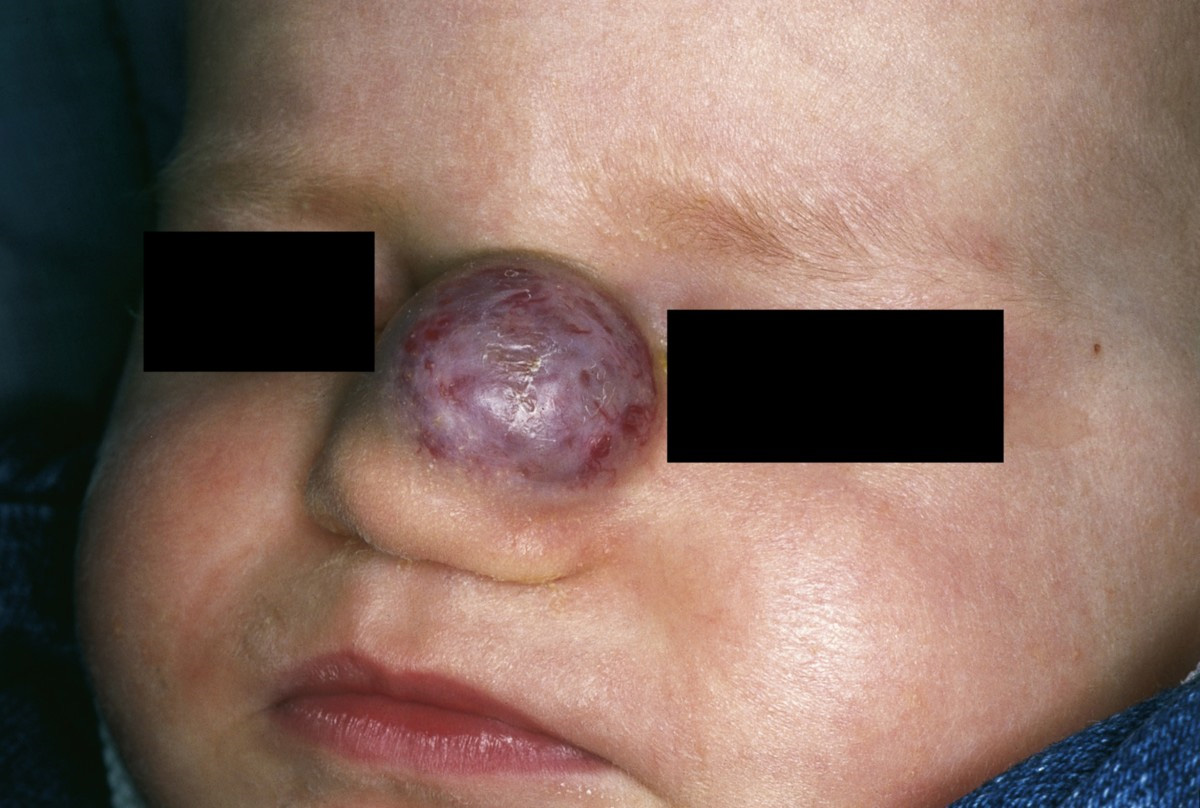
- Any facial haemangioma, not only a large segmental one. The AAP guideline names facial IH in its own right as carrying a risk of permanent scarring and disfigurement, and the nasal tip, the lip and the eyelid are where that risk is highest. This is the gap a size-based list leaves: a moderate lesion on the cheek, nose or lip in a growing infant is a referral, not a watch-and-see
- Ulceration - painful, scarring, and a reason for urgent rather than routine referral
- Five or more cutaneous lesions - consider hepatic haemangiomas and arrange imaging via the specialist team
- A lesion in a baby who is anaemic, has heart failure, or is failing to thrive
Recognise
A bright red, raised, compressible plaque or nodule with a strawberry-like surface when superficial. A deep lesion may show only a bluish swelling with normal overlying skin. Often preceded by a pale patch, a faint telangiectatic mark or a bruise-like area in the first weeks.
The bright strawberry red that gives this lesion its common name is a description of pale skin, and on brown and black skin a superficial haemangioma more often reads as deep red-purple, violaceous or almost black-brown, which is routinely under-recognised and sometimes mistaken for bruising. A deep haemangioma may show no colour change at all. Diagnose by what is tone-independent: a soft compressible swelling, growing visibly week by week in a baby under five months, is a haemangioma until proved otherwise, and the growth history is the finding that matters most. The precursor mark that precedes it - a pale patch or faint telangiectasia - is particularly easy to miss in darker skin, so a parent reporting a mark that is now enlarging deserves to be believed and measured rather than reassured on appearance. Ulceration, which is the commonest complication, is tone-independent. No skin-of-colour series was identified for infantile haemangioma; this draws on general sources.
- The growth curve is the diagnosis. Most rapid and significant growth is between 1 and 3 months of age, and growth is complete by 5 months in most cases
- Soft, compressible, and enlarging - unlike a vascular malformation, which grows with the child
- Often absent or barely visible at birth, then appearing and growing in the first weeks
- Ulceration is the commonest complication needing active treatment, and causes pain and permanent scarring. The commonest lasting consequence, though, is permanent skin change - the AAP guideline puts the risk of scarring and disfigurement above the functional and life-threatening ones, because it affects far more infants
- Up to 5% of infants are affected, so most are ordinary; the job is to pick out the minority that are not
Distribution
Photographs
Mimics
- Benign mechanical petechiae — A new purple mark in a baby raises bruising and safeguarding; a haemangioma is compressible, enlarges over weeks and does not change colour like a bruise
- Molluscum contagiosum — Also discrete papules in an infant, but pearly, umbilicated and multiple, and they do not follow a proliferation curve
- Skin abscess, boils and folliculitis — An ulcerated haemangioma is frequently treated as an abscess or infected lesion; the history of a preceding vascular lesion is what separates them
What to do in the ED
- Establish the age and the growth history, and measure or photograph the lesion. A static lesion and a rapidly growing one need completely different responses, and only the history tells you which this is
- Decide whether any high-risk feature is present - and treat this as a prompt list, not a complete one. Periorbital; beard distribution or any airway concern; any facial lesion, including the nasal tip and lip; large segmental facial or lumbosacral; ulcerated; or five or more lesions. The AAP guideline's own high-risk table is broader than any summary, and includes site and size thresholds this page deliberately does not reproduce because the guideline is paywalled and could not be read in full. If the lesion is facial, large, growing fast, or you are hesitating - refer rather than reassure
- Refer high-risk lesions urgently, and understand why the urgency is real. Growth is fastest between one and three months and is largely over by five months, so the guideline asks for early intervention or referral ideally by 1 month of age. A referral that arrives at six months has missed the window, and many haemangiomas leave permanent skin change behind
- For a stridulous or distressed infant with a beard-distribution lesion, treat as a potential airway emergency and involve ENT and paediatrics immediately - do not refer and discharge
- Treat an ulcerated lesion as painful: analgesia, a non-adherent dressing, and urgent specialist contact
- Do not start propranolol in the ED. It is the systemic drug of choice at 2 to 3 mg/kg/day, usually continued at least six months and often to twelve months of age, but initiation requires cardiovascular assessment and specialist oversight. Topical timolol is an option for selected small, thin, superficial lesions - again, a specialist decision
- For a low-risk lesion, reassure with the natural history and arrange GP or community paediatric follow-up so that growth is actually watched rather than assumed
Disposition
Urgent referral - dermatology, paediatrics or the local vascular anomalies service - for any high-risk feature, and the referral should be measured in days, not the next available clinic. A low-risk lesion in a thriving infant can be managed with GP follow-up and a clear explanation of the growth curve. Airway involvement is an emergency and goes to ENT and paediatrics the same day.
Safety-netting
Local variation
Sources
- Krowchuk DP, Frieden IJ, Mancini AJ, Darrow DH, Blei F, Greene AK, Annam A, Baker CN, Frommelt PC, Hodak A, Pate BM, Pelletier JL, Sandrock D, Weinberg ST, Whelan MA; Subcommittee on the Management of Infantile Hemangiomas. Clinical practice guideline for the management of infantile hemangiomas. Pediatrics 2019;143(1):e20183475
- Mukwende M, Tamony P, Turner M. Mind the Gap: a handbook of clinical signs in black and brown skin. St George's, University of London (2020) - cited for the general principle that erythema and colour change are harder to read on brown and black skin. **It contains no haemangioma content**; no skin-of-colour series for infantile haemangioma was found
- NICE NG240 - rashes may be harder to detect on brown and black skin