Basal cell and squamous cell carcinoma - recognise and refer
Recognise and refer - not a diagnostic tool. The urgencies differ: SCC is a suspected cancer referral, BCC is non-urgent - unless its site or size means delay would matter.
Red flags
- Any skin lesion raising suspicion of squamous cell carcinoma - NICE says consider a suspected cancer pathway referral
- A non-healing ulcer in a chronic wound, burn or scar - consider SCC, and note this pattern is relatively more common in darker skin
- Rapid growth, ulceration, bleeding, or a large lesion. Diameter and depth thresholds are high-risk features in cutaneous SCC staging, not NG12 referral criteria - no figure is quoted here because the UK guideline that carries them has not been read in full. Take the thresholds from it rather than from this page
- Immunosuppression or a transplant recipient with any new or changing lesion - refer promptly
- Perineural symptoms - numbness, tingling or facial weakness near a lesion - suggesting perineural invasion. Like the size thresholds, this is a recognised high-risk feature in SCC staging rather than an NG12 criterion, and it should raise urgency within whichever pathway applies
Recognise
BCC: a pearly or waxy nodule, or an ulcer with a raised rolled edge, with prominent fine blood vessels. SCC: a firm, tender, keratotic or ulcerated nodule that grows over weeks to months.
The pearly translucency and the visible fine telangiectasia that define basal cell carcinoma in pale skin are frequently absent; BCC in brown and black skin is often pigmented, appearing as a dark brown or black nodule or plaque that is readily mistaken for a benign mole or for seborrhoeic keratosis. The raised rolled edge and the central ulceration are structural and remain useful. The more important difference is squamous cell carcinoma, which in Black patients is widely described as arising disproportionately in chronic scars, burns, long-standing ulcers and areas of chronic inflammation rather than in sun-exposed skin. No figure is given for that here because none could be traced to a source read in full, but the practical consequence stands on its own: a non-healing lesion within an old scar or a chronic leg ulcer deserves the same suspicion a sun-exposed lesion would attract in a pale-skinned patient. What is well supported is the outcome - an international expert consensus records that skin cancer in skin of colour often presents at an advanced stage with poorer outcomes, through lower awareness and unfamiliar clinical appearances leading to delayed diagnosis.
- A lesion that does not heal - the most useful single question is how long it has been there
- BCC: NICE describes the typical features as an ulcer with a raised rolled edge, prominent fine blood vessels around a lesion, or a nodule, particularly a pearly or waxy one
- SCC: firmer, faster-growing over weeks to months, often tender, keratotic or ulcerated
- Ask about a non-healing ulcer within a scar, burn or chronic wound
- Immunosuppression, particularly in transplant recipients, greatly increases SCC risk and aggressiveness
Distribution
Photographs
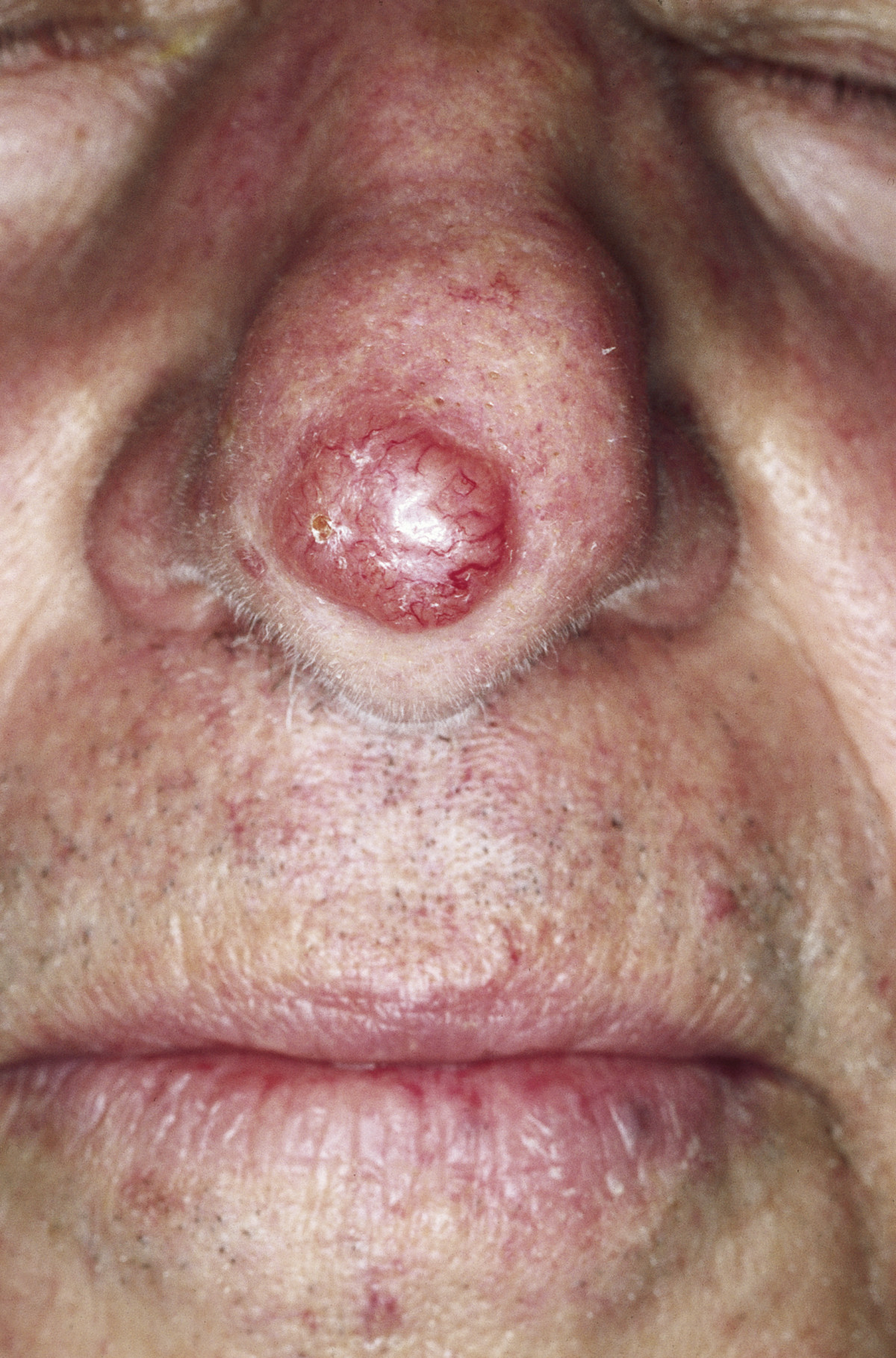
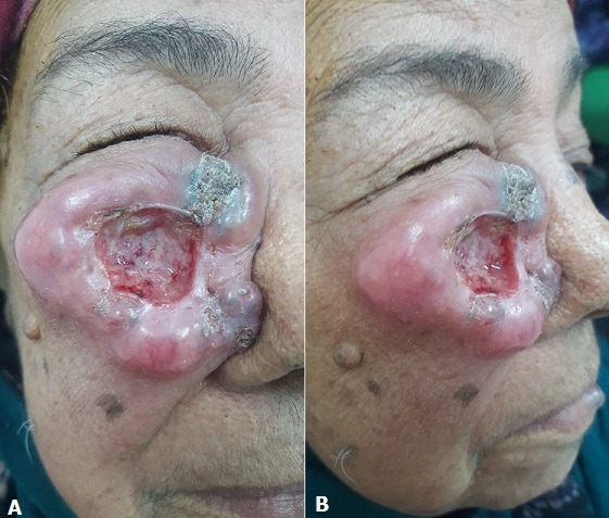
Mimics
- Melanoma - recognise and refer — A pigmented BCC can be mistaken for melanoma and vice versa - both are referred, but on different pathways
- Venous eczema, lipodermatosclerosis and venous leg ulcers — A leg ulcer that is not healing as expected may harbour SCC and warrants biopsy rather than more dressings
- Psoriasis, including guttate — A solitary persistent scaly plaque that does not respond to treatment should be reconsidered, not re-treated
What to do in the ED
- Recognise and refer only. Do not attempt to diagnose, exclude, excise, cauterise or biopsy these lesions in the ED.
- Refer suspected squamous cell carcinoma on a suspected cancer pathway (NG12 1.7.4), and suspected basal cell carcinoma non-urgently (1.7.5) - NICE distinguishes the two explicitly, and conflating them either delays an SCC or floods the pathway
- But NG12 1.7.6 provides the exception, and it matters more than the rule it qualifies: consider a suspected cancer pathway referral for a suspected BCC where there is particular concern that a delay may have a significant impact because of factors such as lesion site or size - a periocular, nasal, auricular or large lesion, for instance. A BCC near the eye is not a non-urgent BCC
- Examine and document the site, size, duration and appearance
- Ask about chronic wounds, scars and burns, and about immunosuppression
- Refer promptly and with a lower threshold in transplant recipients and other immunosuppressed patients
- Do not reassure a patient that a non-healing lesion is benign
- Advise on sun protection without implying that darker skin does not need it
Disposition
Suspected cancer pathway referral for suspected SCC (NG12 1.7.4); non-urgent dermatology referral for suspected BCC (1.7.5) - but use the suspected cancer pathway for a BCC where site or size means a delay would have a significant impact (1.7.6). Lower the threshold and raise the urgency for immunosuppressed patients.
Safety-netting
Local variation
Sources
- NICE NG12 - Suspected cancer: recognition and referral
- Breinholt J et al. Melanoma in Skin of Color: Diagnostic Challenges, Overdiagnosis, and Emerging Tools. Int J Dermatol 2026
- Lim HW et al. International Expert Consensus on Knowledge Gaps in Care for Dermatologic Disorders in Skin of Color. Int J Dermatol 2026
- Keohane SG et al. British Association of Dermatologists guidelines for the management of people with cutaneous squamous cell carcinoma 2020. Br J Dermatol 2021 (not read in full - no PubMed abstract)