Melanoma - recognise and refer
Recognise and refer - this is not a diagnostic tool. The UK referral criterion is the weighted 7-point checklist, not ABCDE.
Red flags
- A weighted 7-point checklist score of 3 or more - suspected cancer pathway referral
- A pigmented or non-pigmented lesion suggesting nodular melanoma - NICE says consider a suspected cancer pathway referral regardless of the checklist score
- A new or changing pigmented band in a single nail, or pigment extending onto the nail fold. Do not attribute nail pigment to an injury and discharge. Patients frequently recall or assume a trauma that did not cause it, and CKS treats associated nail damage as making a new pigmented nail line more suspicious, not less. A subungual haematoma migrates distally and grows out with the nail over weeks; melanoma does not, and pigment on the nail fold is never a haematoma. If you cannot demonstrate it growing out, refer
- Bleeding, ulceration or a rapidly growing lesion
- Any lesion the patient is worried about that you cannot confidently explain - refer rather than reassure
Recognise
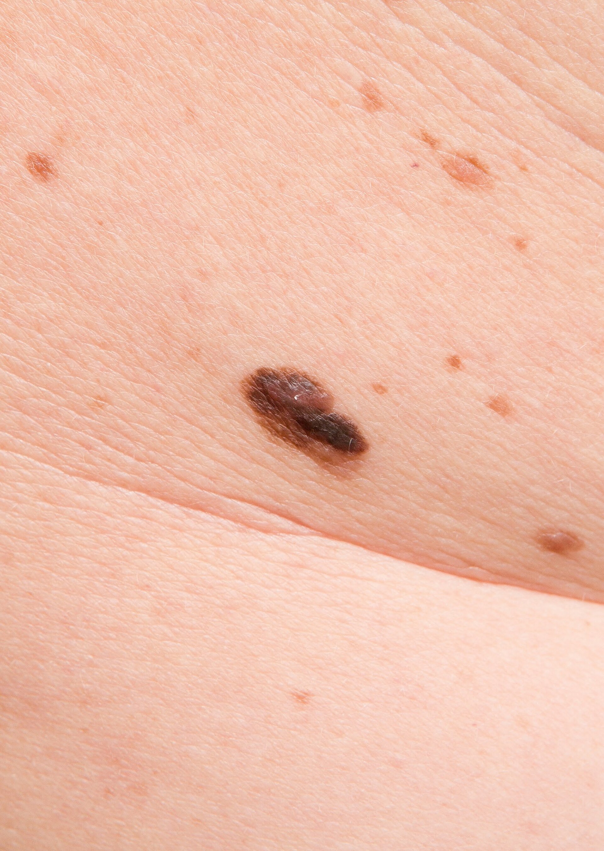
An asymmetric pigmented lesion with an irregular border and variegated colour, changing in size, shape or colour, often on sun-exposed skin; nodular melanoma may be uniform, raised, firm and rapidly growing.
Melanoma is much less common in people with brown and black skin but is diagnosed later and has substantially worse outcomes, and this entry exists because of where it occurs rather than what colour it is. A 2026 review of melanoma in skin of colour, defined there as Fitzpatrick IV to VI, attributes the higher melanoma-specific fatality despite lower incidence to later-stage diagnosis, to differences in subtype distribution, and to systemic barriers to care. Non-Hispanic Black patients in particular have a higher proportion of acral lentiginous and mucosal melanomas, which arise in areas that are not sun-exposed. So the concern is acral lentiginous melanoma, on the palms, soles and nail beds - sites that are not sun-exposed, are routinely not examined, and are not what patients are told to watch. Examine the soles, the palms, between the toes and the nails of anyone presenting with a pigmented lesion. A new, widening or irregular pigmented band in a nail, especially a single nail, and particularly with pigment spreading onto the surrounding nail fold (Hutchinson sign), needs referral - though be aware that benign longitudinal melanonychia is common in people with darker skin, so this is a reason for specialist assessment rather than alarm. Reassurance based on skin tone, or on the absence of sun exposure, is not safe.
- Change is the central concept - in size, shape, colour or sensation
- The UK criterion is NICE's weighted 7-point checklist, with a score of 3 or more triggering a suspected cancer pathway referral
- ABCDE (asymmetry, border, colour, diameter, evolution) is a useful aide-memoire but is not the NICE referral standard
- Nodular melanoma may break all the usual rules - uniform in colour, symmetrical, and simply growing quickly
- A lesion that looks different from the patient's other moles - the ugly duckling
- The checklist was not developed in children and NG12 gives no paediatric variant of it. Melanoma is rare in childhood, but a low score in a child is not a reason to withhold referral - refer a changing, bleeding or rapidly growing lesion on clinical concern alone
- Examine palms, soles and nails, particularly in patients with brown and black skin
Distribution
Photographs
Mimics
- Basal cell and squamous cell carcinoma - recognise and refer — Non-melanoma skin cancers have different referral urgencies - SCC is a suspected cancer pathway referral, BCC is non-urgent
- Molluscum contagiosum — Benign dome-shaped lesions with central umbilication, in clusters, usually in children
What to do in the ED
- This is a recognition and escalation aid only. Do not attempt to diagnose or exclude melanoma from an appearance, and never reassure a patient that a lesion is benign.
- Score the lesion against the weighted 7-point checklist and refer on a suspected cancer pathway if it scores 3 or more
- Refer regardless of score if the lesion suggests nodular melanoma, or if dermoscopy performed by someone competent suggests melanoma
- Examine the palms, soles and nails, particularly in patients with brown or black skin
- Do not excise, shave, biopsy or cauterise a suspicious pigmented lesion in the ED
- Document exactly what you saw, the site, the size, and the checklist score, and make the referral rather than advising the patient to see their GP
- Give the patient a clear account of what happens next and by when
Score
Disposition
Suspected cancer pathway referral for a checklist score of 3 or more, for suspected nodular melanoma, or where dermoscopy suggests melanoma. Make the referral; do not delegate it to the patient.